Glycan labeling strategies and their use in identification and quantification
- PMID: 20225063
- PMCID: PMC2911528
- DOI: 10.1007/s00216-010-3532-z
Glycan labeling strategies and their use in identification and quantification
Abstract
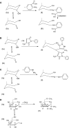
Most methods for the analysis of oligosaccharides from biological sources require a glycan derivatization step: glycans may be derivatized to introduce a chromophore or fluorophore, facilitating detection after chromatographic or electrophoretic separation. Derivatization can also be applied to link charged or hydrophobic groups at the reducing end to enhance glycan separation and mass-spectrometric detection. Moreover, derivatization steps such as permethylation aim at stabilizing sialic acid residues, enhancing mass-spectrometric sensitivity, and supporting detailed structural characterization by (tandem) mass spectrometry. Finally, many glycan labels serve as a linker for oligosaccharide attachment to surfaces or carrier proteins, thereby allowing interaction studies with carbohydrate-binding proteins. In this review, various aspects of glycan labeling, separation, and detection strategies are discussed.
Figures


Similar articles
-
Two-dimensional HPLC separation with reverse-phase-nano-LC-MS/MS for the characterization of glycan pools after labeling with 2-aminobenzamide.Methods Mol Biol. 2009;534:79-91. doi: 10.1007/978-1-59745-022-5_6. Methods Mol Biol. 2009. PMID: 19277534 Review.
-
Capillary electrophoresis/electrospray ion trap mass spectrometry for the analysis of negatively charged derivatized and underivatized glycans.Rapid Commun Mass Spectrom. 2002;16(3):192-200. doi: 10.1002/rcm.564. Rapid Commun Mass Spectrom. 2002. PMID: 11803540
-
Chemoenzymatic method for glycomics: Isolation, identification, and quantitation.Proteomics. 2016 Jan;16(2):241-56. doi: 10.1002/pmic.201500266. Epub 2015 Nov 6. Proteomics. 2016. PMID: 26390280 Free PMC article. Review.
-
Microscale analysis of mucin-type O-glycans by a coordinated fluorophore-assisted carbohydrate electrophoresis and mass spectrometry approach.Electrophoresis. 2003 Feb;24(4):611-21. doi: 10.1002/elps.200390071. Electrophoresis. 2003. PMID: 12601728
-
Comparison of procainamide and 2-aminobenzamide labeling for profiling and identification of glycans by liquid chromatography with fluorescence detection coupled to electrospray ionization-mass spectrometry.Anal Biochem. 2015 Oct 1;486:38-40. doi: 10.1016/j.ab.2015.06.006. Epub 2015 Jun 12. Anal Biochem. 2015. PMID: 26079702
Cited by
-
Systematic comparison of reverse phase and hydrophilic interaction liquid chromatography platforms for the analysis of N-linked glycans.Anal Chem. 2012 Oct 2;84(19):8198-206. doi: 10.1021/ac3012494. Epub 2012 Sep 20. Anal Chem. 2012. PMID: 22954204 Free PMC article.
-
Solid-phase glycan isolation for glycomics analysis.Proteomics Clin Appl. 2012 Dec;6(11-12):596-608. doi: 10.1002/prca.201200045. Proteomics Clin Appl. 2012. PMID: 23090885 Free PMC article. Review.
-
Exopolysaccharide composition and size in Sulfolobus acidocaldarius biofilms.Front Microbiol. 2022 Sep 26;13:982745. doi: 10.3389/fmicb.2022.982745. eCollection 2022. Front Microbiol. 2022. PMID: 36225367 Free PMC article.
-
Harnessing glycomics technologies: integrating structure with function for glycan characterization.Electrophoresis. 2012 Mar;33(5):797-814. doi: 10.1002/elps.201100231. Electrophoresis. 2012. PMID: 22522536 Free PMC article. Review.
-
Purification of Fluorescently Derivatized N-Glycans by Magnetic Iron Nanoparticles.Nanomaterials (Basel). 2019 Oct 17;9(10):1480. doi: 10.3390/nano9101480. Nanomaterials (Basel). 2019. PMID: 31627435 Free PMC article.
References
-
- Parekh RB, Dwek RA, Sutton BJ, Fernandes DL, Leung A, Stanworth D, Rademacher TW, Mizuochi T, Taniguchi T, Matsuta K. Nature. 1985;316:452–457. - PubMed
-
- Peracaula R, Tabares G, Royle L, Harvey DJ, Dwek RA, Rudd PM, De Llorens R. Glycobiology. 2003;13:457–470. - PubMed
-
- Saldova R, Royle L, Radcliffe CM, Abd Hamid UM, Evans R, Arnold JN, Banks RE, Hutson R, Harvey DJ, Antrobus R, Petrescu SM, Dwek RA, Rudd PM. Glycobiology. 2007;17:1344–1356. - PubMed
-
- Jefferis R. Biotechnol Prog. 2005;21:11–16. - PubMed
Publication types
MeSH terms
Substances
LinkOut - more resources
Full Text Sources
Other Literature Sources

