. 2010 Apr 9;6(7):811-7.
doi: 10.1002/smll.200902216.

Gold nanocages as photothermal transducers for cancer treatment

Affiliations

Gold nanocages as photothermal transducers for cancer treatment

Jingyi Chen et al. Small. .

Abstract

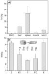
Gold nanocages represent a new class of nanomaterials with compact size and tunable optical properties for biomedical applications. They exhibit strong light absorption in the near-infrared region in which light can penetrate deeply into soft tissue. After PEGylation, the Au nanocages can be passively delivered to tumors in animals. Analysis of tissue distribution for the PEGylated Au nanocages showed that the tumor uptake was 5.7 %ID/g at 96 h post injection. The Au nanocages were found not only on the surface, but also in the core of the tumor. By exposing tumors to a near-infrared diode laser (0.7 W/cm2, CW, λ=808 nm) for 10 min, the photothermal effect of the Au nanocages could selectively destroy tumor tissue with minimum damage to the surrounding healthy tissue. Data from functional [18F]fluorodexoyglucose positron emission tomography revealed a decrease in tumor metabolic activity upon the photothermal treatment. Histological examination identified extensive damage to the nuclei of tumor cells and tumor interstitium.


Figures

Figure 1
Figure 1
UV-vis-NIR spectra showing the LSPR peaks of Au nanocages in different media: PVP-coated nanocages in PBS at pH 7.4 (solid line), PEGylated nanocages in PBS at pH 7.4 (dashed line), and PEGylated nanocages in fetal bovine serum (dotted line). The inset shows a typical TEM image of the Au nanocages with an edge length of 48 nm ± 3.5 nm.
Figure 2
Figure 2
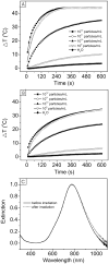
Plots of temperature increase for suspensions of Au nanocages at various concentrations as a function of irradiation time using the diode laser at different power densities: A) 1 W/cm2 and B) 0.5 W/cm2. C) UV-vis-NIR spectra of the Au nanocages in an aqueous solution before (solid line) and after (dashed line) irradiation by the diode laser at a power density of 1 W/cm2 for 10 min.
Figure 3
Figure 3
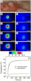
A) Photograph of a tumor-bearing mouse under the photothermal treatment. 100 μL of PEGylated nanocages at a concentration of 9×10¹² particles/mL or saline was administrated intravenously through the tail vein as indicated by an arrow. After the nanocages had been cleared from the circulation (72 h after injection), the tumor on the right frank was irradiated by the diode laser at 0.7 W/cm2 with a beam size indicated by the dashed circle. B–G) Thermographic images of (B–E) nanocage-injected and (F–I) saline-injected tumor-bearing mice at different time points: B, E) 1 min, C, F) 3 min, D, G) 5 min, and E, I) 10 min. J) Plots of average temperature within the tumors (dashed circle) as a function of irradiation time. All scale bars are 1 cm.
Figure 4
Figure 4
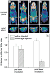
18F-FDG PET/CT co-registered images of mice intravenously administrated with either saline or Au nanocages, followed by laser treatment: A) a saline-injected mouse prior to laser irradiation; B) a nanocage-injected mouse prior to laser irradiation; C) a saline-injected mouse after laser irradiation; and D) a nanocage-injected mouse after laser irradiation. The white arrows indicated the tumors that were exposed to the diode laser at a power density of 0.7 W/cm2 for 10 min. E) A plot showing the ratios of laser-treated tumor (Rt tumor) to non-treated tumor (Lt tumor) 18F-FDG standardized uptake values (SUV, P<0.001).
Figure 5
Figure 5
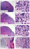
Representative histology images of tumor tissues from the two mice intravenously administrated with saline and Au nanocages, respectively, followed by different treatments: A) tumor from saline-injected mouse with no irradiation; B) tumor from saline-injected mouse with irradiation; C) tumor from nanocage-injected mouse with no irradiation; D) tumor from nanocage-injected mouse with irradiation. The laser treatment was performed by locally irradiated with the diode laser at a power density of 0.7 W/cm2 for 10 min. E–H) The corresponding magnified images of the area in the black boxes shown in the histological photographs (A–D). Tumors from mice treated with nanocages and laser irradiation showed distinctive characteristics of cellular damage, such as abundant pyknosis (arrow), karyorrhexis (open arrow), karyolysis (arrowhead), and interstitial edema (asterisk).
Figure 6
Figure 6
A) Tissue distribution of the PEGylated Au nanocages intravenously administrated (100 μL, with a concentration of 9×10¹² particles/mL) into tumor-bearing mice. The amount of Au in the tissue sample was analyzed by ICP-MS at 96 h post injection. B) Distribution of the PEGylated Au nanocages in the tumor. Note that E, C and EC represent edge, center, and the region between edge and center, respectively. Each data point represents the mean value for n=3 and the bar is the standard deviation for the mean.

References

    1. For reviews: Hu M, Chen J, Li ZY, Au L, Hartland GV, Li X, Marquez M, Xia Y. Chem Soc Rev. 2006;35:1084.Sperling RA, Gil PR, Zhang F, Zanella M, Parak WJ. Chem Soc Rev. 2008;37:1896.Biosselier E, Astrue D. Chem Soc Rev. 2009;38:1759.

    1. Hirsch LR, Stafford RJ, Bankson JA, Sershen SR, Rivera B, Price RE, Hazle JD, Halas NJ, West JL. Proc Natl Acad Sci USA. 2003;100:13549. - PMC - PubMed
    1. Huang X, El-Sayed IH, Qian W, El-Sayed MA. J Am Chem Soc. 2006;128:2115. - PubMed
    1. Chen J, Wang D, Xi J, Au L, Siekkinen A, Warsen A, Li ZY, Zhang H, Xia Y, Li X. Nano Lett. 2007;7:1318. - PMC - PubMed
    1. Melancon MP, Lu W, Yang Z, Zhang R, Cheng Z, Elliot AM, Stafford J, Olson T, Zhang JZ, Li C. Mol Cancer Ther. 2008;7:1730. - PMC - PubMed

Publication types

MeSH terms

LinkOut - more resources