Clinical Trial
. 2010 Feb;67(2):209-20.
doi: 10.1002/ana.21847.

High-frequency electroencephalographic oscillations correlate with outcome of epilepsy surgery

Affiliations
Clinical Trial

High-frequency electroencephalographic oscillations correlate with outcome of epilepsy surgery

Julia Jacobs et al. Ann Neurol. 2010 Feb.

Abstract

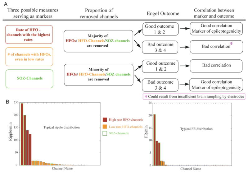
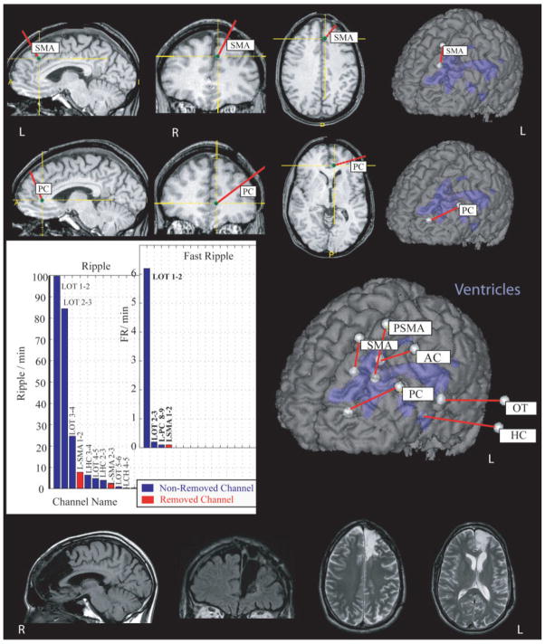
Objective: High-frequency oscillations (HFOs) in the intracerebral electroencephalogram (EEG) have been linked to the seizure onset zone (SOZ). We investigated whether HFOs can delineate epileptogenic areas even outside the SOZ by correlating the resection of HFO-generating areas with surgical outcome.

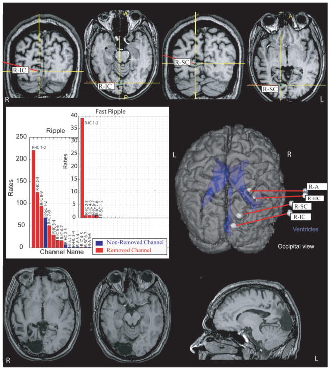
Methods: Twenty patients who underwent a surgical resection for medically intractable epilepsy were studied. All had presurgical intracerebral EEG (500Hz filter and 2,000Hz sampling rate), at least 12-month postsurgical follow-up, and a postsurgical magnetic resonance imaging (MRI). HFOs (ripples, 80-250Hz; fast ripples, >250Hz) were identified visually during 5 to 10 minutes of slow-wave sleep. Rates and extent of HFOs and interictal spikes in resected versus nonresected areas, assessed on postsurgical MRIs, were compared with surgical outcome (Engel's classification). We also evaluated the predictive value of removing the SOZ in terms of surgical outcome.

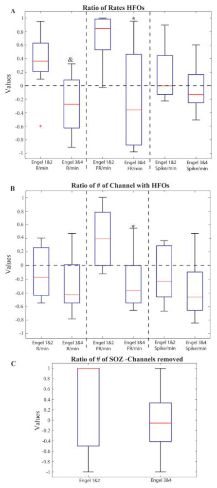
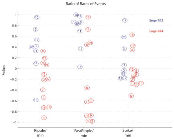
Results: The mean duration of follow-up was 22.7 months. Eight patients had good (Engel classes 1 and 2) and 12 poor (classes 3 and 4) surgical outcomes. Patients with a good outcome had a significantly larger proportion of HFO-generating areas removed than patients with a poor outcome. No such difference was seen for spike-generating regions or the SOZ.

Interpretation: The correlation between removal of HFO-generating areas and good surgical outcome indicates that HFOs could be used as a marker of epileptogenicity and may be more accurate than spike-generating areas or the SOZ. In patients in whom the majority of HFO-generating tissue remained, a poor surgical outcome occurred.


Figures

FIGURE 1
FIGURE 1
This figure demonstrates our hypothesis. (A) The diagram illustrates the assumed relationship between the removal of the majority of measured events and the surgical outcome. Our hypothesis does not assume that all patients with a poor outcome must have remaining high-frequency oscillation (HFO)-generating regions. It might be that the majority of recorded HFOs are removed, but HFOs are generated in brain areas in which there are no electrodes. (B) Histograms in which channels are ranked according to event rates. The 2 graphs illustrate a typical distribution of ripples (left), some contacts having very high rates and many contacts having low rates, and fast ripples (FRs) (right), with similar numbers of contacts with high and low rates. In all patients, the distribution of FRs was restricted to much fewer channels than that of ripples. SOZ = seizure onset zone.
FIGURE 2
FIGURE 2
Overview of the results of the study. (A) Ratio of event rates in the removed areas to rates in the non-removed areas for patients with good versus bad outcome (see formula in text). Regions generating high rates of ripples (R) and fast ripples (FRs) where more likely to have been removed in patients with good outcome. (B) Ratio of the number of contacts with events to the number of contacts without any event in patients with good versus poor surgical outcomes (see formula in text). Only the number of contacts carrying FRs showed a significant difference. (C) The ratio of removed versus nonremoved seizure onset zone (SOZ) contacts is compared for the 2 outcome groups. No significant difference was seen. *Significantly different from Engel classes 1 and 2 (p = 0.04). &Significantly different from Engel classes 1 and 2 (p = 0.008). Red lines signify median. HFO = high-frequency oscillation. [Color figure can be viewed in the online issue, which is available at www.interscience.wiley.com.]
FIGURE 3
FIGURE 3
Same data as in Figure 2A, but showing all patients. All patients with good outcome are in blue and patients with poor outcome in red circles. A number identifies each patient, so that it is possible to follow which marker (ripples on the left, fast ripples [FRs] in the middle, spikes on the right) is indicative of the outcome in each patient. Values >0 indicate that the majority of high-frequency oscillations (HFOs) are removed and patients should therefore have a good outcome (blue circles). One patient did not have any FRs (#7). The chart shows that, except for patient #15, all patients with good outcome had the majority of HFOs removed (values >0). This result can be stated differently: all patients with most HFOs remaining (values <0) had a poor outcome (except patient #15). We also notice that for patients with poor outcome, all but 1 (#19) had the majority of ripples or FRs remaining.
FIGURE 4
FIGURE 4
Pre- and postsurgical magnetic resonance imaging (MRI) of patient #10 as an example of a patient with removal of most of high-frequency oscillation-generating tissue and a good outcome. The postsurgical MRI clearly shows that the areas around electrode R-IC were removed, whereas the tissue around the inner contacts of R-SC remained untouched. The axial view of the postsurgical MRI also demonstrates the electrode traces of R-A and R-HC. The graph illustrates the rates of ripples/fast ripples in contacts that were removed (red) and those that remained (blue). Only channels that had events are shown; ripples are found in more channels than fast ripples. A = amygdala; HC = hippocampus; IC = infracalcarine; L = left; R = right; SC = supracalcarine.
FIGURE 5
FIGURE 5
Patient #4 underwent a resection in the left frontal lobe with removal of the brain areas around the electrodes in the left anterior cingulate (L-AC) and left supplementary motor area (L-SMA). High-frequency oscillations were most frequently seen in the contacts placed in the occipitotemporal junction. The patient had a poor outcome, with reduced but persisting complex partial seizures. HC = hippocampus; OT= occipitotemporal junction; PC = posterior cingulate; PSMA = posterior SMA; R = right.

References

    1. Schuele SU, Lüders HO. Intractable epilepsy: management and therapeutic alternatives. Lancet Neurol. 2008;7:514–524. - PubMed
    1. Diehl B, Lüders HO. Temporal lobe epilepsy: when are invasive recordings needed? Epilepsia. 2000;41(suppl 3):61–74. - PubMed
    1. Rosenow F, Lüders H. Presurgical evaluation of epilepsy. Brain. 2001;124(pt 9):1683–1700. - PubMed
    1. Boling W, Aghakhani Y, Andermann F, Sziklas V, Olivier A. Surgical treatment of independent bitemporal lobe epilepsy defined by invasive recordings. J Neurol Neurosurg Psychiatry. 2009;80:533–538. - PubMed
    1. Prasad A, Pacia SV, Vazquez B, et al. Extent of ictal origin in mesial temporal sclerosis patients monitored with subdural intracranial electrodes predicts outcome. Clin Neurophysiol. 2003;20:243–248. - PubMed

Publication types

MeSH terms