. 2010 Feb;67(2):221-9.
doi: 10.1002/ana.21871.

Microemboli may link spreading depression, migraine aura, and patent foramen ovale

Affiliations

Microemboli may link spreading depression, migraine aura, and patent foramen ovale

Ala Nozari et al. Ann Neurol. 2010 Feb.

Abstract

Objective: Patent foramen ovale and pulmonary arteriovenous shunts are associated with serious complications such as cerebral emboli, stroke, and migraine with aura. The pathophysiological mechanisms that link these conditions are unknown. We aimed to establish a mechanism linking microembolization to migraine aura in an experimental animal model.

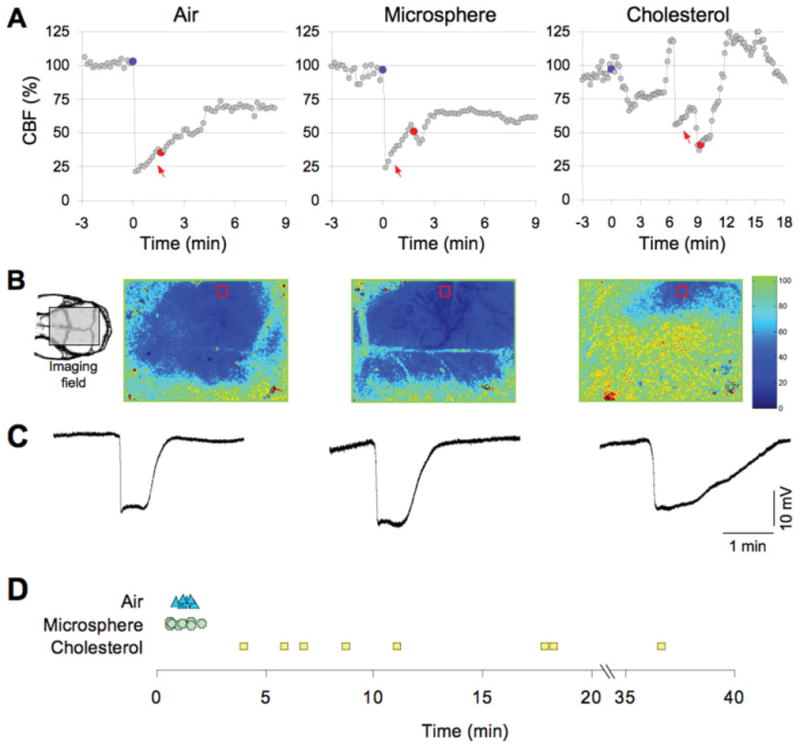
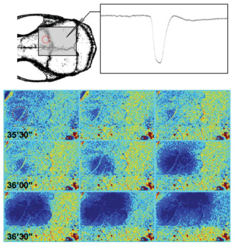
Methods: We introduced particulate or air microemboli into the carotid circulation in mice to determine whether transient microvascular occlusion, insufficient to cause infarcts, triggered cortical spreading depression (CSD), a propagating slow depolarization that underlies migraine aura.

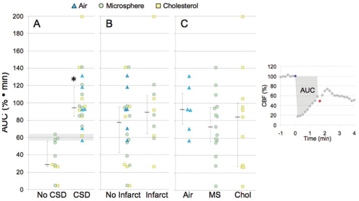
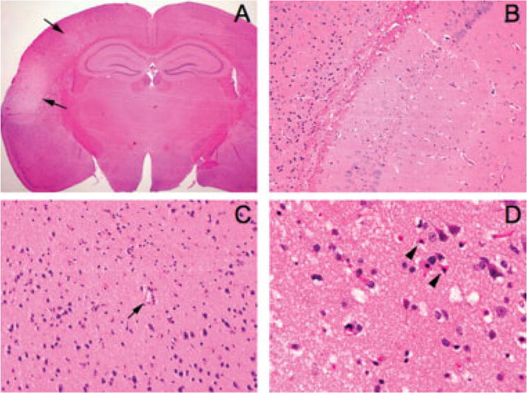
Results: Air microemboli reliably triggered CSD without causing infarction. Polystyrene microspheres (10 microm) or cholesterol crystals (<70 microm) triggered CSD in 16 of 28 mice, with 60% of the mice (40% of those with CSD) showing no infarcts or inflammation on detailed histological analysis of serial brain sections. No evidence of injury was detected on magnetic resonance imaging examination (9.4T; T2 weighted) in 14 of 15 selected animals. The occurrence of CSD appeared to be related to the magnitude and duration of flow reduction, with a triggering mechanism that depended on decreased brain perfusion but not sustained tissue damage.

Interpretation: In a mouse model, microemboli triggered CSD, often without causing microinfarction. Paradoxical embolization then may link cardiac and extracardiac right-to-left shunts to migraine aura. If translatable to humans, a subset of migraine auras may belong to a spectrum of hypoperfusion disorders along with transient ischemic attacks and silent infarcts.


Figures

FIGURE 1
FIGURE 1
Microembolic hypoperfusion triggers cortical spreading depression (CSD). (A) Representative tracings show the time course of cerebral blood flow (CBF) reduction after intracarotid air, microsphere, or cholesterol injection (blue circles). Both air and microspheres abruptly but transiently decreased CBF to ≤25% of baseline, whereas the hypoperfusion after cholesterol microemboli was less predictable and fluctuated over time (note the compressed time scale for the cholesterol data). CSD (red circles) occurred within a few minutes after air or microsphere embolization, but was more delayed after cholesterol microemboli. CSDs were detected by the characteristic slow extracellular direct current (DC) shift shown in C and by a propagating wave of hypoperfusion. (B) Full-field images of CBF taken at the denoted time point between microembolization (red arrows in A) and CSD onset showing the spatial extent of hypoperfusion. Air and microspheres reduced CBF within the ipsilateral middle cerebral artery (MCA) and bilateral anterior cerebral artery territories, whereas cholesterol-induced hypoperfusion was more restricted to ipsilateral MCA branches. Red squares show the regions of interest within which the CBF time courses (shown in A) were quantified for each type of microemboli. The orientation of the imaging field over the mouse skull is shown in the inset (left). The inset shows the position of the rectangular imaging field. (C) Extracellular DC potential shifts characteristic of CSD were triggered by intracarotid microembolization with air (left), microsphere (middle), or cholesterol (right), and recorded by intracortical glass micropipettes within the ipsilateral MCA territory simultaneously with the laser speckle flowmetry. (D) The timing of CSD after air (triangles), microsphere (crosses), and cholesterol microemboli (squares) is shown for each animal. Both air and microspheres evoked a CSD within a few minutes after microembolization, whereas the onset of CSD was significantly delayed after cholesterol microemboli.
FIGURE 2
FIGURE 2
Cortical spreading depression (CSD) originates from a hypoperfused cortical focus. Representative time-lapse laser speckle images of relative cerebral blood flow changes taken approximately 35 minutes after cholesterol microembolization show the initiation of a CSD from a frontal cortical focus of hypoperfusion (red circle). A CSD was initiated from this ischemic focus after approximately 30 seconds (36′00‴), and propagated throughout the ipsilateral cortex (centrifugally spreading blue hypoperfusion wave), confirmed by concurrent electrophysiological recordings using a glass micropipette (inset). Images were acquired at 0.1Hz. Laser speckle imaging field and electrode positions are shown on the upper left drawing.
FIGURE 3
FIGURE 3
Ischemic burden and its relationship to cortical spreading depression (CSD) and infarct occurrence. Ischemic burden (area under the curve [AUC]) was calculated by measuring the area under the cerebral blood flow (CBF) curve between the injection of microemboli (blue circle) and the onset of CSD (red circle), as shown in the inset on the right. (A) The occurrence of CSD was associated with significantly higher AUC values than experiments without CSD; 60% · min (horizontal shaded area) was the AUC threshold for evoking CSD. (B) AUC values did not differ significantly between the animals with or without infarcts, regardless of the embolic particle. (C) The AUC values did not differ between the 3 types of microemboli. Individual data points as well as median (25–75% range) are given for each group. *p < 0.01 vs no CSD; Mann-Whitney rank sum test. MS = microsphere; Chol = = cholesteol.
FIGURE 4
FIGURE 4
Histopathology of microembolic infarcts. (A) A representative coronal section showing 2 cortical infarcts (arrows) 72 hours after injection of microspheres (10μm diameter, 20,000 particles; original magnification, ×12.5). (B) Although the majority of infarcts were in the ipsilateral cortex, the hippocampal formation also showed microinfarcts in a few brains (original magnification, ×200). (C) Most cortical infarcts were centered around a small blood vessel (arrow) (original magnification, ×200). (D) High-power image of a representative cortical microinfarct demonstrating the characteristic shrunken, eosinophilic neurons with pyknotic nuclei (arrowheads) adjacent to intact neurons at the infarct edge (original magnification, ×400).

References

    1. Lipton RB, Stewart WF, Diamond S, et al. Prevalence and burden of migraine in the United States: data from the American Migraine Study II. Headache. 2001;41:646–657. - PubMed
    1. Henrich JB, Horwitz RI. A controlled study of ischemic stroke risk in migraine patients. J Clin Epidemiol. 1989;42:773–780. - PubMed
    1. Etminan M, Takkouche B, Isorna FC, Samii A. Risk of ischaemic stroke in people with migraine: systematic review and meta-analysis of observational studies. BMJ. 2005;330:63. - PMC - PubMed
    1. Moskowitz MA. The neurobiology of vascular head pain. Ann Neurol. 1984;16:157–168. - PubMed
    1. Leao AAP. Spreading depression of activity in the cerebral cortex. J Neurophysiol. 1944;7:359–390. - PubMed

Publication types

MeSH terms