. 2010 Apr 20;49(15):3351-65.
doi: 10.1021/bi100198y.

Anthranilate-activating modules from fungal nonribosomal peptide assembly lines

Affiliations

Anthranilate-activating modules from fungal nonribosomal peptide assembly lines

Brian D Ames et al. Biochemistry. .

Abstract

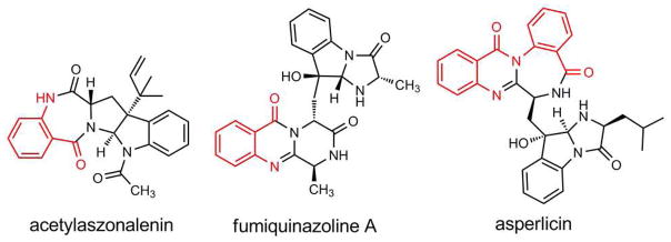
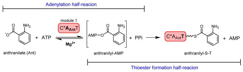
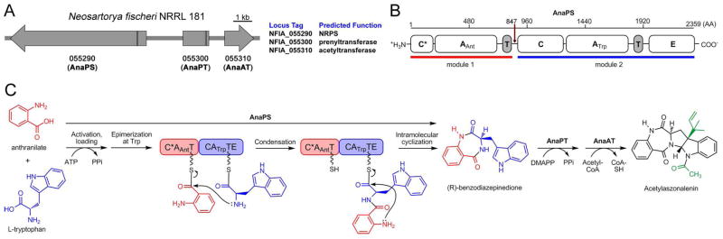
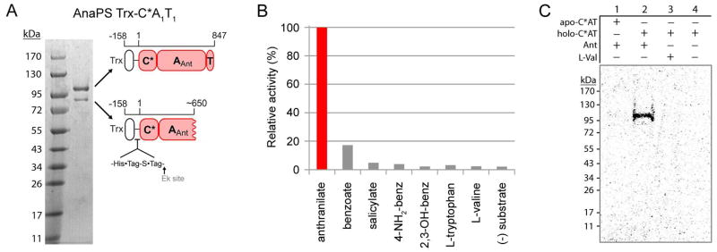
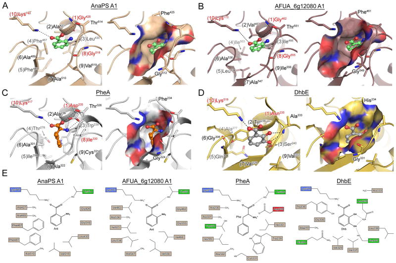
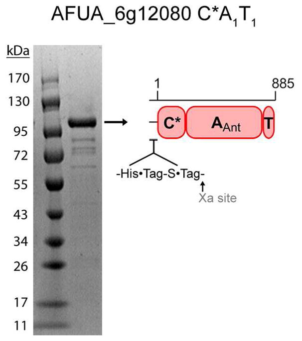
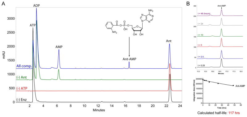
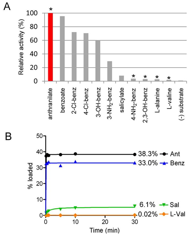
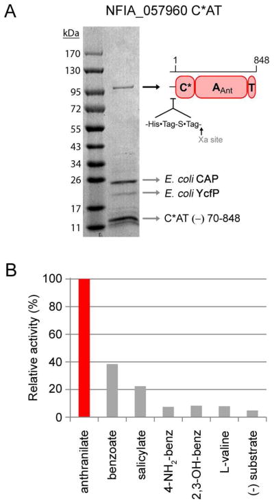
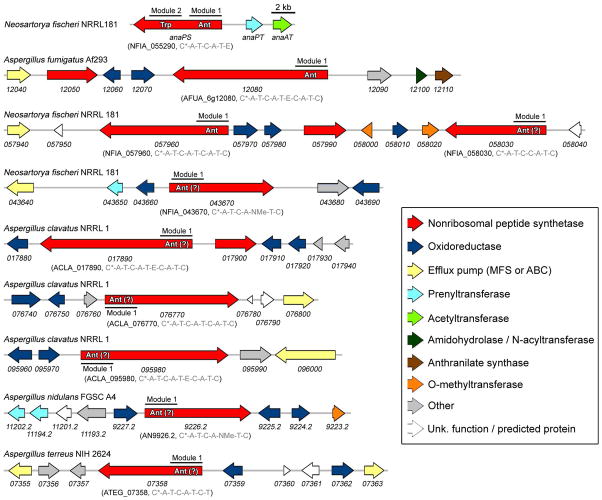
Fungal natural products containing benzodiazepinone- and quinazolinone-fused ring systems can be assembled by nonribosomal peptide synthetases (NRPS) using the conformationally restricted beta-amino acid anthranilate as one of the key building blocks. We validated that the first module of the acetylaszonalenin synthetase of Neosartorya fischeri NRRL 181 activates anthranilate to anthranilyl-AMP. With this as a starting point, we then used bioinformatic predictions about fungal adenylation domain selectivities to identify and confirm an anthranilate-activating module in the fumiquinazoline A producer Aspergillus fumigatus Af293 as well as a second anthranilate-activating NRPS in N. fischeri. This establishes an anthranilate adenylation domain code for fungal NRPS and should facilitate detection and cloning of gene clusters for benzodiazepine- and quinazoline-containing polycyclic alkaloids with a wide range of biological activities.


Figures

Figure 1
Figure 1
Examples of fungal natural products that utilize anthranilic acid as a building block (Ant derived portion highlighted in red).
Figure 2
Figure 2
Scheme for the chemical steps of anthranilic acid activation (by adenylation) and anthranilyl transfer (by thiolation) catalyzed by the A- and T-domains of fungal NRPS module 1 constructs characterized in this work. The wavy line of the anthranilyl-S-T intermediate corresponds to the phosphopantetheine (PPT) prosthetic group.
Figure 3
Figure 3
Acetylaszonalenin biosynthesis from Neosartorya fischeri NRRL 181. (A) Annotated three-gene cluster. Vertical black lines in the Orfs demarcate introns. (B) Domain organization of the bimodular NRPS AnaPS, illustrating predicted domain boundaries as determined from bioinformatics analysis. Red arrow marks the “cutoff” point used for cloning module 1 C*AT. (C) Proposed route of acetylaszonalelin biosynthesis. Previous work has validated the conversion of (R)-benzodiazepinedione to acetylaszonalenin by AnaAT and AnaPT, and in this work we validate the activation and loading of anthranilate by AnaPS module 1. The loading and epimerization of Trp, and the condensation and cyclization reactions are hypothetical.
Figure 4
Figure 4
Purification and characterization of AnaPS module 1. (A) Purity of the thioredoxin fusion protein of AnaPS module 1 (Trx-C*A1T1) post Ni-NTA chromatography and concentration-induced protein precipitation. Cartoon representation is used to indicate the identity and domain-composition of the two prominent bands present in the gel as determined by peptide mass fingerprinting. (B) ATP-[32P]PPi exchange assay data obtained for Trx-C*AT. 100% relative activity for anthranilate-dependent exchange corresponds to 6200 CPM. (C) Autoradiograph of SDS-PAGE gel illustrating the covalent loading of [carboxy-14C]anthranilate onto the holo-T-domain of Trx-C*AT.
Figure 5
Figure 5
Homology models and crystal structures of NRPS adenylation domains. (A) and (B) are homology models of the proposed Ant-activating A-domains of AnaPS module 1 and AFUG_6g12080 module 1, respectively (PheA used as template). Anthranilic acid was manually docked into the binding pocket based on the PheA and DhbE structures and is rendered as green ball-and-stick. (C) Crystal structure of PheA (PDB ID: 1AMU) bound to AMP and L-Phe (orange ball-and-stick). (D) Crystal structure of DhbE (PDB ID: 1MDB) bound to 2,3-dihyhydroxybenzoyl-AMP (2,3-DHB rendered as grey ball-and-stick). For (A)-(D), left image shows specificity-determining residues as sticks, with potential hydrogen bond and/or ionic interactions drawn as dashed lines. Numbers in parenthesis preceding the residue label indicate residue position according to the 10AA specificity code. Right image presents a molecular surface representation of the substrate binding pocket colored according to electrostatic properties (blue = positive, red = negative), and includes two residues which are conserved among A-domains as Gly and Phe/His/Tyr. (E) 2D representation of the substrate selectivity residues for panels (A)–(D).
Figure 6
Figure 6
Purified recombinant AFUA_6g12080 C*A1T1 following heterologous expression in E. coli and Ni-affinity chromatography. The identity of the indicated band as the full-length protein was confirmed by peptide mass fingerprinting.
Figure 7
Figure 7
HPLC-based determination of anthranilyl-AMP formation by AFUA_6g12080 module 1 C*AT and stability of Ant-AMP following release from enzyme. (A) Overlay of HPLC chromatograms (251 nm detection) obtained from 20 μL injections of control (black, red, and green traces) and experimental (blue trace) reactions monitoring the in vitro adenylation activity of C*AT with anthranilic acid as substrate. All reactions contained 50 mM HEPES (pH 7.4), 10 mM MgCl2, and 1 mM DTT; and varied combinations of 10 μM C*AT (“Enz”), 5 mM ATP, and 1 mM Ant as indicated. The chemical identity of peaks was determined by running authentic standards and high-resolution mass spectrometry. (B) The stability of Ant-AMP in reaction buffer following release from enzyme. Top image, overlay of HPLC chromatograms (251 nm detection) obtained at different timepoints post microcentrifugal filtration (10K molecular weight cutoff) of a sample prepared similar to the “All components” reaction presented in panel (A). Bottom image, quantification of Ant-AMP peak area for each timepoint used for determination of Ant-AMP stability in terms of calculated half-life.
Figure 8
Figure 8
Biochemical characterization of AFUA_6g12080 module 1 C*AT. (A) ATP-[32P]PPi exchange data obtained for the adenylation activity of AFUA_6g12080 C*AT using various aryl and L-amino acids. Asterisks mark molecules which are known components of fungal primary metabolism (see Discussion). (B) Timecourse comparison for the in vitro loading of 14C-labeled anthranilate, benzoate, salicylate, and L-valine onto the holo-T-domain of C*AT.
Figure 9
Figure 9
Purification and characterization of NFIA_057960 module 1 C*AT. (A) Purity of the C*AT protein following Ni-NTA, anion-exchange, and gel-filtration chromatography. The identity of the ~100 kDa band as full-length C*AT protein, and the identities of the low-molecular-weight impurities indicated by arrows were determined by mass fingerprinting (CAP, catabolite gene activator protein; YcfP, conserved hypothetical protein; C*AT (−) 70-848, fragment of full-length protein missing residues ≈ 70-848). (B) Adenylation activity of C*AT with a panel of aryl-acids assessed by ATP-[32P]PPi exchange assay. L-valine is provided as a negative control.
Figure 10
Figure 10
Organization of fungal biosynthetic gene clusters containing, or proposed to contain, anthranilic acid-activating nonribosomal peptide synthetases. The locus tag and NRPS domain organization (from N- to C-terminus) is provided in parenthesis. The minimum domains constituting a single NRPS module include C*/C-A-T. Additional domains sometimes present as part of a single module are an E-domain (epimerization), or an NMe-domain (N-methylation). The N-terminal C* domains of NFIA_058030, NFIA_043670, and AN9226.2 are about one-third the size of the C* domains of the other NRPSs (which are predicted to be defined by ~200 residues).

Similar articles

Cited by

References

    1. D’Yakonov A, Telezhenetskaya M. Quinazoline alkaloids in nature. Chem Nat Compd. 1997;33:221–267.
    1. Finking R, Marahiel MA. Biosynthesis of nonribosomal peptides. Annu Rev Microbiol. 2004;58:453–488. - PubMed
    1. Sattely ES, Fischbach MA, Walsh CT. Total biosynthesis: in vitro reconstitution of polyketide and nonribosomal peptide pathways. Nat Prod Rep. 2008;25:757–793. - PubMed
    1. Yeulet SE, Mantle PG. Biosynthesis of auranthine by Penicillium aurantiogriseum. FEMS Microbiol Lett. 1987;41:207–210.
    1. Houck DR, et al. On the biosynthesis of asperlicin and the directed biosynthesis of analogs in Aspergillus alliaceus. J Antibiot (Tokyo) 1988;41:882–891. - PubMed

Publication types