

TDP-43 and ubiquitinated cytoplasmic aggregates in sporadic ALS are low frequency and widely distributed in the lower motor neuron columns independent of disease spread
- PMID: 20225928
- PMCID: PMC4981144
- DOI: 10.3109/17482961003602363
TDP-43 and ubiquitinated cytoplasmic aggregates in sporadic ALS are low frequency and widely distributed in the lower motor neuron columns independent of disease spread
Abstract
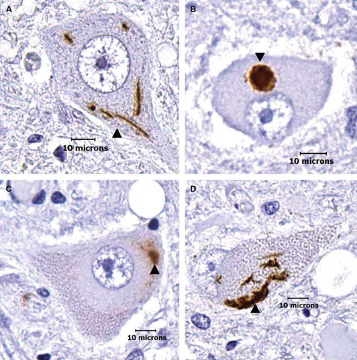
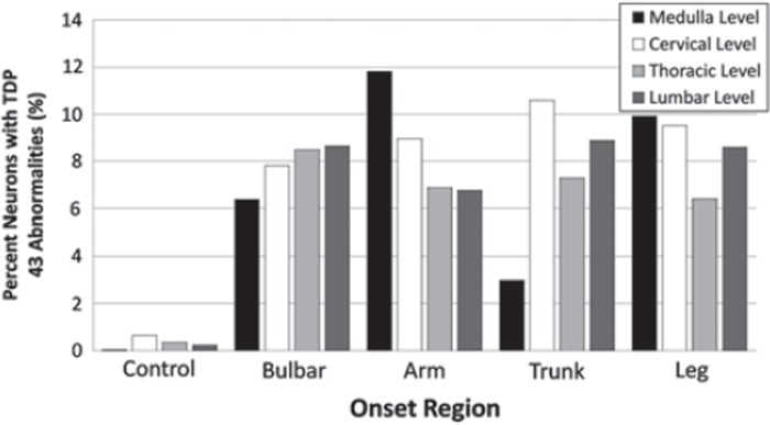
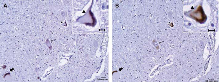
Ubiqitinated and TDP-43 immunoreactive cytoplasmic aggregates are hallmark features of ALS molecular pathology. Since clinically most ALS begins focally and advances contiguously, it is important to characterize their distribution. Our objective was to determine the extent and distribution of TDP-43 immunoreactive aggregates in the lower motor neuron columns as a function of disease onset, and to correlate ubiquitinated with TDP-43 aggregates in the lumbar region. We examined TDP-43 cytoplasmic aggregates at four separate neuraxis levels - hypoglossal nucleus and cervical, thoracic, and lumbar anterior horns - in five controls and 20 sporadic ALS nervous systems from patients whose disease began in various sites, i.e. five bulbar, five arm, five trunk, and five leg onsets. We correlated ubiquitinated to TDP-43 aggregates on adjacent histological sections for the lumbar regions. We found that TDP-43 cytoplasmic aggregates are seen in about 8% of motor neurons but there is marked variability between nervous systems, ranging from 0.4% to 20.6%. The aggregates are uniformly distributed within individual nervous systems. There is no obvious correlation between site of disease onset and rate of spread. Almost all ubiquitinated aggregates correlate to TDP-43 aggregates. Thus, TDP-43 immunoreactive cytoplasmic aggregates have a low overall average frequency that does not correlate with either disease course or clinical spread and is the prime ubiquitinated protein.
Figures
References
-
- Leigh PN, Anderton BH, Dodson A, Gallo JM, Swash M, Power DM. Ubiquitin deposits in anterior horn cells in motor neuron disease. Neuroscience Letters. 1988;93:197–203. - PubMed
-
- Neumann M, Sampathu DM, Kwong LK, Truax AC, Micsenyi MC, Chou TT. Ubiquitinated TDP-43 in frontotemporal lobar degeneration and amyotrophic lateral sclerosis. Science. 2006;314:130–3. - PubMed
-
- Arai T, Hasegawa M, Akiyama H, Ikeda K, Nonaka T, Mori H. TDP-43 is a component of ubiquitin-positive tau-negative inclusions in frontotemporal lobar degeneration and amyotrophic lateral sclerosis. Biochemical and Biophysical Research Communications. 2006;351:602–11. - PubMed
-
- Mackenzie IR, Bigio EH, Ince PG, Geser F, Neumann M, Cairns NJ. Pathological TDP-43 distinguishes sporadic amyotrophic lateral sclerosis from amyotrophic lateral sclerosis with SOD1 mutations. Ann Neurol. 2007;61:427–34. - PubMed
-
- Robertson J, Sanelli T, Xiao S, Yang W, Horne P, Hammond R. Lack of TDP-43 abnormalities in mutant SOD1 transgenic mice shows disparity with ALS. Neurosci Lett. 2007;420:128–32. - PubMed
Publication types
MeSH terms
Substances
LinkOut - more resources
Full Text Sources
Medical
Miscellaneous