. 2010 Mar 12:11:21.
doi: 10.1186/1471-2199-11-21.

Regulation of TFIIIB during F9 cell differentiation

Affiliations

Regulation of TFIIIB during F9 cell differentiation

Dimitris Athineos et al. BMC Mol Biol. .

Abstract

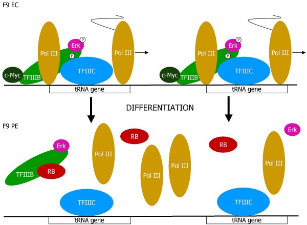
Background: Differentiation of F9 embryonal carcinoma (EC) cells into parietal endoderm (PE) provides a tractable model system for studying molecular events during early and inaccessible stages of murine development. PE formation is accompanied by extensive changes in gene expression both in vivo and in culture. One of the most dramatic is the ~10-fold decrease in transcriptional output by RNA polymerase (pol) III. This has been attributed to changes in activity of TFIIIB, a factor that is necessary and sufficient to recruit pol III to promoters. The goal of this study was to identify molecular changes that can account for the low activity of TFIIIB following F9 cell differentiation.

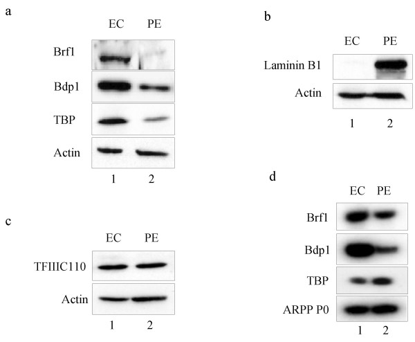
Results: Three essential subunits of TFIIIB decrease in abundance as F9 cells differentiate; these are Brf1 and Bdp1, which are pol III-specific, and TBP, which is also used by pols I and II. The decreased levels of Brf1 and Bdp1 proteins can be explained by reduced expression of the corresponding mRNAs. However, this is not the case for TBP, which is regulated post-transcriptionally. In proliferating cells, pol III transcription is stimulated by the proto-oncogene product c-Myc and the mitogen-activated protein kinase Erk, both of which bind to TFIIIB. However, c-Myc levels fall during differentiation and Erk becomes inactive through dephosphorylation. The diminished abundance of TFIIIB is therefore likely to be compounded by changes to these positive regulators that are required for its full activity. In addition, PE cells have elevated levels of the retinoblastoma protein RB, which is known to bind and repress TFIIIB.

Conclusion: The low activity of TFIIIB in PE can be attributed to a combination of changes, any one of which could be sufficient to inhibit pol III transcription. Declining levels of essential TFIIIB subunits and of activators that are required for maximal TFIIIB activity are accompanied by an increase in a potent repressor of TFIIIB. These events provide fail-safe guarantees to ensure that pol III output is appropriate to the diminished metabolic requirements of terminally differentiated cells.


Figures

Figure 1
Figure 1
Expression of exogenous Brf1 in F9 cell stable transfectants. Western blot of whole cell extract (20 μg) from untransfected F9 cells (lane 1), Brf1.F9 cells (lane 2) and vec.F9 cells (lane 3) probed with antibodies 128 against Brf1 and C-11 against actin, as indicated.
Figure 2
Figure 2
Stable expression of Brf1 in F9 cells does not stimulate proliferation. a, b Proliferation curves showing numbers of viable Brf1.F9 (blue) and vec.F9 (red) cells, as determined by trypan blue staining, each day after plating 104 undifferentiated EC cells (a) or differentiated PE cells (b). Counts were taken in triplicate and the results are presented as averages of three independent experiments +/- standard deviation. c RT-PCR analysis to compare expression of tRNAiMet and tRNALeu, and mRNAs encoding Brf1 and ARPP P0 in vec.F9 and Brf1.F9 cells, as indicated.
Figure 3
Figure 3
Stable expression of Brf1 in F9 cells does not prevent down-regulation of tRNA gene transcription following differentiation. a Western blot of whole cell extract (50 μg) from untransfected F9 cells (lanes 1 and 2) and Brf1.F9 cells (lanes 3 and 4) before (EC) or after (PE) differentiation. Blots were probed with antibodies F-7 against HA and C-11 against actin, as indicated. b Quantitative comparison of tRNALeu gene transcription in vitro using extracts (20 μg) of Brf1.F9 and untransfected F9 cells before (EC) and after (PE) differentiation. Graph shows mean from three independent experiments +/- standard deviation. c Representative example of one of the transcription assays quantified in Fig. 3b.
Figure 4
Figure 4
Differentiation of F9 EC cells into PE is accompanied by specific decreases in expression of Brf1, Bdp1 and TBP proteins, as well as Brf1 and Bdp1 mRNAs. a Western blot of whole cell extract (50 μg) from untransfected F9 cells before (EC) or after (PE) differentiation and probed with antibodies against Brf1, Bdp1, TBP and actin, as indicated. b Western blot of whole cell extract (20 μg) from untransfected F9 cells before (EC) or after (PE) differentiation and probed with antibodies against laminin B1 and actin, as indicated. c Western blot of whole cell extract (50 μg) from untransfected F9 cells before (EC) or after (PE) differentiation and probed with antibodies against TFIIIC110 and actin, as indicated. d RT-PCR analysis to compare expression of mRNAs encoding Brf1, Bdp1, TBP and ARPP P0 in undifferentiated (EC) and differentiated (PE) F9 cells, as indicated.
Figure 5
Figure 5
Differentiation of F9 EC cells into PE is accompanied by specific changes in regulators of TFIIIB activity. a Western blot of whole cell extract (20 μg) of undifferentiated (EC) and differentiated (PE) F9 cells using antibodies against RB, p107, p130 and TFIIB, as indicated. b Western blot of whole cell extract (20 μg) of undifferentiated (EC) and differentiated (PE) F9 cells using antibodies against c-Myc, Max, TFIIB and actin, as indicated. c Western blot of whole cell extract (20 μg) of undifferentiated (EC) and differentiated (PE) F9 cells using antibodies against total Erk and active (phosphorylated) Erk, as well as TFIIB, as indicated.
Figure 6
Figure 6
Model of changes to the pol III transcription machinery that accompany differentiation of F9 EC cells into PE. In EC cells, tRNA genes are transcribed very actively, reflecting the availability of TFIIIB, c-Myc and activated Erk. Differentiation is accompanied by decreased levels of TFIIIB and c-Myc, as well as inactivation of Erk and induction of RB. PE cells contain relatively little TFIIIB, which is deprived of activators and liable to inhibition by RB; as a consequence, transcription of tRNA genes is severely restricted.

Similar articles

Cited by

References

    1. White RJ, Stott D, Rigby PWJ. Regulation of RNA polymerase III transcription in response to F9 embryonal carcinoma stem cell differentiation. Cell. 1989;59:1081–1092. doi: 10.1016/0092-8674(89)90764-2. - DOI - PubMed
    1. Alzuherri HM, White RJ. Regulation of a TATA-binding protein-associated factor during cellular differentiation. J Biol Chem. 1998;273:17166–17171. doi: 10.1074/jbc.273.27.17166. - DOI - PubMed
    1. Perletti L, Kopf E, Carre L, Davidson I. Coordinate regulation of RARgamma2, TBP, and TAFII 135 by targeted proteolysis during retinoic acid-induced differentiation of F9 embryonal carcinoma cells. BMC Mol Biol. 2001;2:4. doi: 10.1186/1471-2199-2-4. - DOI - PMC - PubMed
    1. Meiβner W, Ahlers A, Seifart KH. The activity of transcription factor PBP, which binds to the proximal sequence element of mammalian U6 genes, is regulated during differentiation of F9 cells. Mol Cell Biol. 1995;15:5888–5897. - PMC - PubMed
    1. Meissner W, Thomae R, Seifart KH. The activity of transcription factor IIIC1 is impaired during differentiation of F9 cells. J Biol Chem. 2002;277:7148–7156. doi: 10.1074/jbc.M108721200. - DOI - PubMed

Publication types

MeSH terms

LinkOut - more resources