. 2010 Mar 12:11:170.
doi: 10.1186/1471-2164-11-170.

The RST and PARP-like domain containing SRO protein family: analysis of protein structure, function and conservation in land plants

Affiliations

The RST and PARP-like domain containing SRO protein family: analysis of protein structure, function and conservation in land plants

Pinja Jaspers et al. BMC Genomics. .

Abstract

Background: The SROs (SIMILAR TO RCD-ONE) are a group of plant-specific proteins which have important functions in stress adaptation and development. They contain the catalytic core of the poly(ADP-ribose) polymerase (PARP) domain and a C-terminal RST (RCD-SRO-TAF4) domain. In addition to these domains, several, but not all, SROs contain an N-terminal WWE domain.

Results: SROs are present in all analyzed land plants and sequence analysis differentiates between two structurally distinct groups; cryptogams and monocots possess only group I SROs whereas eudicots also contain group II. Group I SROs possess an N-terminal WWE domain (PS50918) but the WWE domain is lacking in group II SROs. Group I domain structure is widely represented in organisms as distant as humans (for example, HsPARP11). We propose a unified nomenclature for the SRO family. The SROs are able to interact with transcription factors through the C-terminal RST domain but themselves are generally not regulated at the transcriptional level. The most conserved feature of the SROs is the catalytic core of the poly(ADP-ribose) polymerase (PS51059) domain. However, bioinformatic analysis of the SRO PARP domain fold-structure and biochemical assays of AtRCD1 suggested that SROs do not possess ADP-ribosyl transferase activity.

Conclusions: The SROs are a highly conserved family of plant specific proteins. Sequence analysis of the RST domain implicates a highly preserved protein structure in that region. This might have implications for functional conservation. We suggest that, despite the presence of the catalytic core of the PARP domain, the SROs do not possess ADP-ribosyl transferase activity. Nevertheless, the function of SROs is critical for plants and might be related to transcription factor regulation and complex formation.


Figures
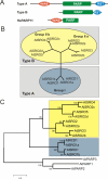
Figure 1
Figure 1
Structural classes and evolutionary relationships of RCD1 and RCD1-like proteins. (A) Schematic diagram depicting the protein structure of A. thaliana RCD1/SRO protein family members representing the two structural classes, type A (AtRCD1) and B (AtSRO2). RCD1 and all SROs possess a poly(ADP-ribose) polymerase (PARP) catalytic region (PS51059) and a C-terminal RST (RCD1-SRO-TAF4; PF12174) domain. Additionally, the presence (type A) or absence (type B) of a WWE domain (PS50918) differentiates between the two structural classes. Human HsPARP11 exhibits similar domain composition to the Arabidopsis SROs containing a WWE domain and the PARP domain. It is representative of the five human WWE-PARPs although the remaining four have additional conserved domains. (B) A. thaliana and A. lyrata SROs clustered in three groups in an unrooted Neighbour-joining tree. AtRCD1 and AtSRO1 and the A. lyrata orthologs AlSRO1a and AlSRO1b formed group I, which structurally belongs to type A. AtSRO2 and AtSRO3 and the A. lyrata orthologs AlSRO2a and AlSRO2b formed group IIa. AtSRO4 and AtSRO5 and the A. lyrata orthologs AlSRO2c and AlSRO2d form group IIb. All members of group II belong to structural type B. (C) Neighbour-joining tree of the A. thaliana and A. lyrata SROs rooted the A. thaliana PARPs (AtPARP1, 2 and 3). The SRO proteins clustered together and form a monophyletic group while AtPARP1, 2 and 3 clustered together to form a single outgroup for the SRO protein family.
Figure 2
Figure 2
Transcript profile of SRO family genes. Bootstrapped Bayesian hierarchical clustering of the A. thaliana SRO family genes under various stresses compared to normal growth conditions. The stress data sets were downloaded from public databases (see Methods for complete description of the method and the data). Red and green indicate increased or decreased expression compared to untreated plants, respectively. The intensity of the colors is proportional to the absolute value of the fold difference.
Figure 3
Figure 3
qPCR analysis of SRO family genes. Steady state transcript levels of A. thaliana SRO family genes were investigated by qPCR. Relative gene expression under light stress, salt stress, and exposure to O3 and in the rcd1-2 mutant is shown compared to Col-0 wildtype plants grown under normal conditions. Red indicates elevated and green decreased expression. Black indicates unaltered transcript levels or in the case of AtSRO4 not reproducible (NR). Numbers indicate relative fold-change ratios. All experiments were repeated three times, one representative experiment is shown.
Figure 4
Figure 4
Real-time quantitative PCR analysis of the sro5-2 mutant. The expression of 13 genes which were most differentially expressed in non-stressed sro5-2 mutant plants according to microarray results (data not shown) was re-examined by qPCR. Red indicates elevated and black unaltered transcript levels compared to Col-0 wildtype plants. Numbers indicate relative fold-change ratios. All experiments were repeated three times, one representative experiment is shown.
Figure 5
Figure 5
SRO Orthologs in Sequenced Plant Genomes. All SRO sequences used for analyses are listed with names according to the proposed nomenclature and their original identifiers. The length of the proteins in amino acids (AAs; size) and the presence (+) or absence (-) of potential conserved domains (WWE PS50918, PARP PS51059, RST PF12174) are indicated. Proteins predicted to lack domains because they are not full length are indicated (#). Domains present but with low statistical support are indicated with (‡). Data source: NCBI (National Center for Bioinformatics, http://www.ncbi.nlm.nih.gov/). Additional web resources are listed below: PZ, Phytozome http://www.phytozome.net/; TAIR, the Arabidopsis Information Resource http://www.arabidopsis.org/; JGI, Joint Genome Initiative (Poplar: http://genome.jgi-psf.org/Poptr1_1/Poptr1_1.home.html; Physcomitrella: http://genome.jgi-psf.org/Phypa1_1/Phypa1_1.home.html; Selaginella: http://genome.jgi-psf.org/Selmo1/Selmo1.home.html); CVI, Craig Venter Insititute http://castorbean.jcvi.org/; BDB, Brachypodium database http://www.brachypodium.org/; RGADB, Rice Genome Annotation database http://rice.plantbiology.msu.edu/. Two SROs from Brachypodium, Bradi2g10720.1 and Bradi1g01340.1, were only present as very short and incomplete predictions, and thus could not be assigned to any group.
Figure 6
Figure 6
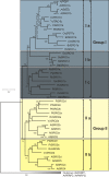
Neighbour-joining tree of the PARP domains of the plant SRO protein family. The PARP domains of the SRO proteins from the sequenced genomes of A. thaliana, A. lyrata, Vitis vinifera, Ricinus communis, Populus trichocarpa, Oryza sativa ssp. japonica, Brachypodium distachyon, Physcomitrella patens and Selaginella moellendorffi were identified and aligned. Subsequently, a Neighbour-joining phylogenetic tree was constructed using MEGA4. AtPARP1, 2 and 3 were used as outgroups. Plant SROs could be classified into two groups. Group I contained SROs from all included plant species and could be further divided into three subgroups (Ia, Ib and Ic) according to the C-terminal RST domain. Most SROs in group I belonged to structural type A. The members of group II (a and b) without exception belonged to structural type B.
Figure 7
Figure 7
The RST domain of the plant SRO protein family contains a strongly conserved amino acid pattern. (A) Domain structure of AtRCD1 and TAF4s from multiple species (Saccharomyces cerevisiae, Schizosaccharomyces pombe, Homo sapiens, Drosophila melanogaster). All TAF4s have the conserved TAF4 superfamily domain (TAF4; PF05236). Yeast TAF4s lack an N-terminal extension while metazoan TAF4s have an extension bearing an ETO domain (ETO/TAFH domain; PF07531), which is a known transcription factor-recruitment domain [43]. Plant TAF4s also have an N-terminal extension that lacks the ETO domain but bears the structurally unrelated plant-specific RST (RCD1-SRO-TAF4; PF12174) domain. TAF4 RST has not been tested for TF interaction, however, the RST domain from AtRCD1 is required for interaction with multiple TFs. AtRCD1 also bears PARP-like (PS51059) and WWE (PS50918) domains. (B) The C-terminal RST domain of the different groups and subgroups (Ia, Ib, Ic, IIa, IIb) of the plant SRO protein family were aligned using ClustalW and Boxshade. Consensus sequences for each group or subgroup are depicted in bold characters and marked according to similarity: conserved (*), strong similarty (:), weak similarity (.) using Boxshade. Under the sequence, alternatives for AAs are shown. AAs with similar chemical properties are indicated using colored bars. Green indicates polar, non-charged, non-aliphatic residues. Blue indicates the most hydrophobic AAs. Red indicates positively charged AAs. Magenta highlights acidic residues. Orange shows glycine and brown indicates tyrosine.
Figure 8
Figure 8
The RST domain of AtRCD1 is required for the TF interactions. The C-terminus of AtRCD1 was truncated to determine the minimum protein length capable of interacting with TFs. The dark gray horizontal bars above the AtRCD1 protein sequence denote the different constructs. Green background indicates interaction and gray background the lack of it. Yeast spots from each interaction test are depicted in the panel on the right. The ClustalW alignment of AtRCD1, AtSRO1 and AtSRO5 is included for comparison of the RST structure in different proteins. Highlighted AAs in the protein sequences are as in figure 7.
Figure 9
Figure 9
AtSRO5 interacts with transcription factors. Transcription factors interacting with AtSRO5 in a pairwise interaction test against the REGIA TF collection. FL: Full length AtRCD1. PCT: AtRCD1 construct lacking the WWE domain. + interaction observed, - interaction not observed.
Figure 10
Figure 10
Subcellular localization of AtSRO5. The AtSRO5-GFP fusion protein localized to several dot-like structures in the nucleus in A. thaliana seedlings (panels A-C). As comparison, the mitochondrial localization marker line mt-yk (panels D-F) and the nuclear and cytoplasmic localization of YFP protein (panels G-I) are shown. Panels A, D and G display the fluorescent signal, panels B, E and H the light micrograph and panels C, F and I the two overlaid. Scale bar 5 μm; arrows in B, C, H and I indicate the nucleus.
Figure 11
Figure 11
Conserved active site fold structure of the PARP domain. Fold-assisted AA alignments of the PARP catalytic core from (A) human PARP1 (HsPARP1), A. thaliana PARP-1 and -2 (AtPARP1, AtPARP2) and RCD1 (AtRCD1) and (B) A. thaliana RCD1 and SROs (AtSRO1-5). Consensus of conserved (*) and similar (: and .) AAs and conserved folds (α-helix or β-sheet) are indicated below the alignments. Additionally folds are shaded in the alignment with grey (α-helix) or yellow (β-sheet) backgrounds. Conserved ADP-ribosyl transferase catalytic triad, composed of three AAs at the C-terminus of β-sheet 1, middle of β-sheet 2 and N-terminal end of β-sheet 5, is indicated by turquoise background shading and an (‡) above the alignment. Alignments were performed with T-Coffee at EMBL-EBI http://www.ebi.ac.uk/Tools and hand-adjusted according to fold predictions performed with Psipred in the Phyre search [53]. AAs were color-coded according to their biochemical properties as in http://www.ebi.ac.uk/Tools/t-coffee/help.html#color.
Figure 12
Figure 12
AtRCD1 does not bind NAD and does not have ADP-ribosylation activity. Biochemical analysis of NAD binding and ART activity of AtRCD1. (A) NAD binding analysis: Autoradiography image of a SDS-PAGE gel showing proteins labeled with [32P-NAD] upon UV irradiation. SAD-A, RCD1-GST, PCT-GST or GST were incubated with 0.6 μM of [32P-NAD] in absence or presence of 0.6 mM of unlabeled NAD under UV light (see Methods). (B) Picture of the SDS-PAGE gel shown in (A) stained with Coomassie Brilliant Blue. Positions of RCD1-GST and PCT-GST are marked on panels (A) and (B) with arrows; asterisks mark the position of the DnaK protein. (C) ART activity analysis: Autoradiography image of SDS-PAGE gel showing poly-ADP-ribosylation of proteins in presence of [32P-NAD]. HsPARP1, RCD1-GST or PCT-GST in concentration 200 nM were incubated with 1.3 μM [32P-NAD] (see Methods) in absence or presence of 3 μg of histones. (D) Picture of the SDS-PAGE gel shown in (C) stained with Coomassie Brilliant Blue. The 70 kDa band represents BSA used as a carrier for protein precipitation. All panels: Unlabeled NAD was used in the competition experiments. Molecular weight marker sizes (kDa) are indicated on the left side of each panel. The experiment was repeated three times with similar results, one representative experiment is shown.

Similar articles

Cited by

References

    1. Jaspers P, Blomster T, Brosché M, Salojärvi J, Ahlfors R, Vainonen JP, Reddy RA, Immink R, Angenent G, Turck F, Overmyer K, Kangasjärvi J. Unequally redundant RCD1 and SRO1 mediate stress and developmental responses and interact with transcription factors. Plant J. 2009;60(2):268–79. doi: 10.1111/j.1365-313X.2009.03951.x. - DOI - PubMed
    1. Teotia S, Lamb RS. The paralogous genes RADICAL-INDUCED CELL DEATH 1 and SIMILAR TO RCD ONE 1 have partially redundant functions during Arabidopsis thaliana development. Plant Physiol. 2009;151(1):180–98. doi: 10.1104/pp.109.142786. - DOI - PMC - PubMed
    1. Belles-Boix E, Babiychuk E, Van Montagu M, Inzé D, Kushnir S. CEO1, a new protein from Arabidopsis thaliana, protects yeast against oxidative damage. FEBS Lett. 2000;482(1-2):19–24. doi: 10.1016/S0014-5793(00)02016-0. - DOI - PubMed
    1. Overmyer K, Tuominen H, Kettunen R, Betz C, Langebartels C, Sandermann H, Kangasjärvi J. Ozone-sensitive Arabidopsis rcd1 mutant reveals opposite roles for ethylene and jasmonate signaling pathways in regulating superoxide-dependent cell death. Plant Cell. 2000;12(10):1849–62. doi: 10.1105/tpc.12.10.1849. - DOI - PMC - PubMed
    1. Ahlfors R, Lång S, Overmyer K, Jaspers P, Brosché M, Tauriainen A, Kollist H, Tuominen H, Belles-Boix E, Piippo M, Inzé D, Palva ET, Kangasjärvi J. Arabidopsis RADICAL-INDUCED CELL DEATH1 belongs to the WWE protein-protein interaction domain protein family and modulates abscisic acid, ethylene, and methyl jasmonate responses. Plant Cell. 2004;16(7):1925–37. doi: 10.1105/tpc.021832. - DOI - PMC - PubMed

Publication types