

Binding and activation of host plasminogen on the surface of Francisella tularensis
- PMID: 20226053
- PMCID: PMC2848021
- DOI: 10.1186/1471-2180-10-76
Binding and activation of host plasminogen on the surface of Francisella tularensis
Abstract
Background: Francisella tularensis (FT) is a gram-negative facultative intracellular coccobacillus and is the causal agent of a life-threatening zoonotic disease known as tularemia. Although FT preferentially infects phagocytic cells of the host, recent evidence suggests that a significant number of bacteria can be found extracellularly in the plasma fraction of the blood during active infection. This observation suggests that the interaction between FT and host plasma components may play an important role in survival and dissemination of the bacterium during the course of infection. Plasminogen (PLG) is a protein zymogen that is found in abundance in the blood of mammalian hosts. A number of both gram-positive and gram-negative bacterial pathogens have the ability to bind to PLG, giving them a survival advantage by increasing their ability to penetrate extracellular matrices and cross tissue barriers.
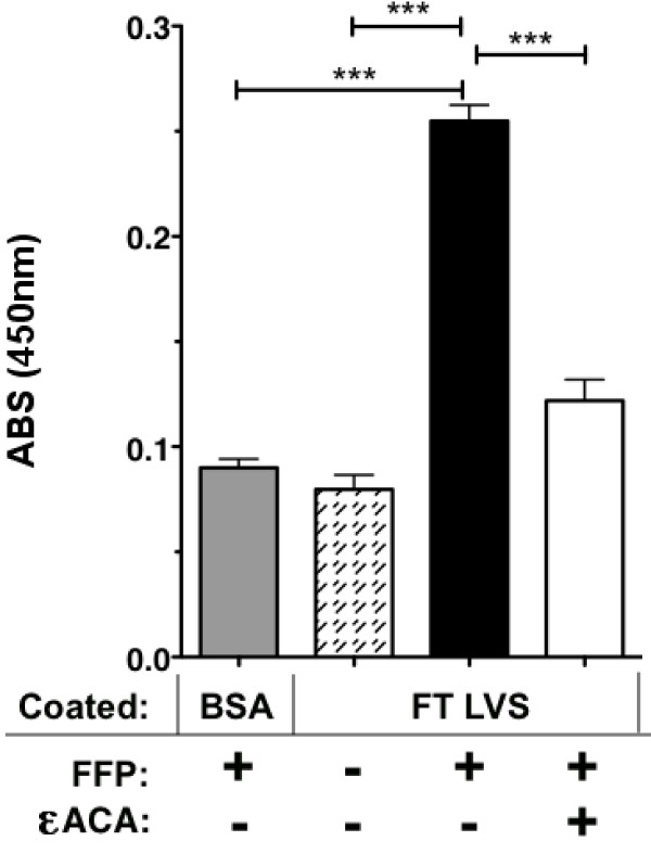
Results: We show that PLG binds to the surface of FT and that surface-bound PLG can be activated to plasmin in the presence of tissue PLG activator in vitro. In addition, using Far-Western blotting assays coupled with proteomic analyses of FT outer membrane preparations, we have identified several putative PLG-binding proteins of FT.
Conclusions: The ability of FT to acquire surface bound PLG that can be activated on its surface may be an important virulence mechanism that results in an increase in initial infectivity, survival, and/or dissemination of this bacterium in vivo.
Figures
Similar articles
-
TolC and EmrA1 contribute to Francisella novicida multidrug resistance and modulation of host cell death.J Bacteriol. 2024 Sep 19;206(9):e0024624. doi: 10.1128/jb.00246-24. Epub 2024 Aug 28. J Bacteriol. 2024. PMID: 39194223 Free PMC article.
-
Deletion of the Major Facilitator Superfamily Transporter fptB Alters Host Cell Interactions and Attenuates Virulence of Type A Francisella tularensis.Infect Immun. 2018 Feb 20;86(3):e00832-17. doi: 10.1128/IAI.00832-17. Print 2018 Mar. Infect Immun. 2018. PMID: 29311235 Free PMC article.
-
Plasminogen binding proteins and plasmin generation on the surface of Leptospira spp.: the contribution to the bacteria-host interactions.J Biomed Biotechnol. 2012;2012:758513. doi: 10.1155/2012/758513. Epub 2012 Oct 15. J Biomed Biotechnol. 2012. PMID: 23118516 Free PMC article.
-
Exploitation of plasmin(ogen) by bacterial pathogens of veterinary significance.Vet Microbiol. 2015 Jul 9;178(1-2):1-13. doi: 10.1016/j.vetmic.2015.04.008. Epub 2015 Apr 17. Vet Microbiol. 2015. PMID: 25937317 Review.
-
Proteins involved in Francisella tularensis survival and replication inside macrophages.Future Microbiol. 2012 Nov;7(11):1255-68. doi: 10.2217/fmb.12.103. Future Microbiol. 2012. PMID: 23075445 Review.
Cited by
-
XFEL and NMR Structures of Francisella Lipoprotein Reveal Conformational Space of Drug Target against Tularemia.Structure. 2020 May 5;28(5):540-547.e3. doi: 10.1016/j.str.2020.02.005. Epub 2020 Mar 5. Structure. 2020. PMID: 32142641 Free PMC article.
-
De novo design of high-affinity miniprotein binders targeting Francisella tularensis virulence factor.bioRxiv [Preprint]. 2025 Jul 5:2025.07.02.662053. doi: 10.1101/2025.07.02.662053. bioRxiv. 2025. PMID: 40631088 Free PMC article. Preprint.
-
Subversion of host recognition and defense systems by Francisella spp.Microbiol Mol Biol Rev. 2012 Jun;76(2):383-404. doi: 10.1128/MMBR.05027-11. Microbiol Mol Biol Rev. 2012. PMID: 22688817 Free PMC article. Review.
-
Shotgun Immunoproteomic Approach for the Discovery of Linear B-Cell Epitopes in Biothreat Agents Francisella tularensis and Burkholderia pseudomallei.Front Immunol. 2021 Sep 29;12:716676. doi: 10.3389/fimmu.2021.716676. eCollection 2021. Front Immunol. 2021. PMID: 34659206 Free PMC article.
-
Host-adaptation of Francisella tularensis alters the bacterium's surface-carbohydrates to hinder effectors of innate and adaptive immunity.PLoS One. 2011;6(7):e22335. doi: 10.1371/journal.pone.0022335. Epub 2011 Jul 22. PLoS One. 2011. PMID: 21799828 Free PMC article.
References
-
- Siret V, Barataud D, Prat M, Vaillant V, Ansart S, Le Coustumier A, Vaissaire J, Raffi F, Garre M, Capek I. An outbreak of airborne tularaemia in France, August 2004. Euro Surveill. 2006;11(2):58–60. - PubMed
-
- Syrjala H, Kujala P, Myllyla V, Salminen A. Airborne transmission of tularemia in farmers. Scand J Infect Dis. 1985;17(4):371–375. - PubMed
Publication types
MeSH terms
Substances
Grants and funding
LinkOut - more resources
Full Text Sources
Other Literature Sources
Miscellaneous
