. 2010 Feb 1;1(1):8.
doi: 10.1186/1759-8753-1-8.

Immunity of replicating Mu to self-integration: a novel mechanism employing MuB protein

Affiliations

Immunity of replicating Mu to self-integration: a novel mechanism employing MuB protein

Jun Ge et al. Mob DNA. .

Abstract

We describe a new immunity mechanism that protects actively replicating/transposing Mu from self-integration. We show that this mechanism is distinct from the established cis-immunity mechanism, which operates by removal of MuB protein from DNA adjacent to Mu ends. MuB normally promotes integration into DNA to which it is bound, hence its removal prevents use of this DNA as target. Contrary to what might be expected from a cis-immunity mechanism, strong binding of MuB was observed throughout the Mu genome. We also show that the cis-immunity mechanism is apparently functional outside Mu ends, but that the level of protection offered by this mechanism is insufficient to explain the protection seen inside Mu. Thus, both strong binding of MuB inside and poor immunity outside Mu testify to a mechanism of immunity distinct from cis-immunity, which we call 'Mu genome immunity'. MuB has the potential to coat the Mu genome and prevent auto-integration as previously observed in vitro on synthetic A/T-only DNA, where strong MuB binding occluded the entire bound region from Mu insertions. The existence of two rival immunity mechanisms within and outside the Mu genome, both employing MuB, suggests that the replicating Mu genome must be segregated into an independent chromosomal domain. We propose a model for how formation of a 'Mu domain' may be aided by specific Mu sequences and nucleoid-associated proteins, promoting polymerization of MuB on the genome to form a barrier against self-integration.


Figures
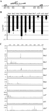
Figure 1
Figure 1
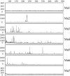
Quantification and profile of Mu insertions within Mu in vivo. (a) Experimental strategy. A common MuR primer (labeled* with 6FAM for experiments in (c)), anneals within the right end or attR of Mu. Primers annealing to seven different locations within Mu (Mu1 to Mu7) were each paired with MuR in PCR reactions to measure (b) the frequency and (c) the site of Mu insertions packaged in phage particles. Wavy lines indicate host DNA linked to Mu ends in packaged phage heads. (b) Real-time PCR reactions were performed using Mu DNA purified from phage prepared after induction of strain SJG3. Ct values are inversely proportional to the amount of nucleic acid of interest in the sample. Log2 of the relative insertion frequency (RIF) values were derived from Ct differences between samples and input Mu DNA control; primer pairs annealing within region 4 served as controls for input DNA. Primers hybridizing to E. coli genes known to be hot (yidP), cold (rfaS) and average (ahpF) Mu insertion targets were also paired with MuR as controls. Primer efficiencies were calculated as described in Methods. The data are an average of three technical repeats. (c) Profile of Mu insertions within the targets monitored in (b). PCR reactions were as in (b), except that MuR was labeled with the fluorescent primer 6FAM. The reactions were subjected to FLA. Numbers on the X axis refer to nucleotides. The intensity of the fluorescent signal reflects the frequency of insertion at a particular site and is represented by arbitrary numbers on the Y axis.
Figure 2
Figure 2
Immunity of the linear Mu genome in vitro. (a) Transposition reactions were set up by incubating MuA, MuB and HU proteins with the mini-Mu plasmid pSP104 as donor and either linear Mu genome or pUC19 as target, as described in Methods. Lane 1, cleaved Type I complex assembled on pSP104; lanes 2 to 4, Type II or strand transfer reactions with: pSP104 as donor and either pUC19 (lane 2) or Mu (lane 3) as target or Mu as donor and pUC19 as target (lane 4); lanes 5 to 7, control substrates without added proteins. (b) Map of pSP104 showing restriction enzyme sites used for linearization and their position with respect to the attL and attR Mu ends. (c) Transposition reactions with linear mini-Mu as target. Reaction conditions were as in (a), except that Type I complexes were first assembled on pSP104 and added to indicated targets in a second step. Lane1, Type I reaction; Lane 2 to 7, Type II reactions. Lanes 8 to 13, DNA controls without added proteins. Restriction enzyme shown in the B panel are abbreviated to H, X and B. L = linear. O = open circular; S = supercoiled; Type I = cleaved complex; Type II = strand transfer complex; Type II (intra) = intramolecular strand transfer complexes.
Figure 3
Figure 3
Profile of Mu insertions within the Mu genome in vitro. Reactions were as described in Figure 1c, except that the template for PCR was the in vitro Type II reaction shown in Figure 2, lane 3.
Figure 4
Figure 4
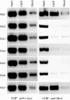
ChIP reactions probing binding of c-myc-MuB and MuA on the Mu genome during Mu replication. ChIP samples were prepared with either anti-c-myc antibody or anti-MuA antibody using strain SJG3 as described in Methods. Binding on the seven different segments of Mu genome shown in Figure 1a was tested by regular PCR amplification. Input = fragmented whole genome DNA; ChIP = DNA in ChIP samples; Mock = DNA recovered without addition of specific antibody during the ChIP procedure.
Figure 5
Figure 5
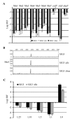
qPCR of DNA isolated by ChIP using anti-c-myc-MuB antibody after induction of Mu replication. (a) Location of DNA segments within and outside the Mu prophage. The Mu insertion is in malF, whose direction of transcription on the E. coli genome is indicated by arrows. The L0 region spans 45 to 328 bp from the beginning of attR, whereas the R0 region spans 89 to 343 bp from the beginning of attL. L1 to L25 and R1 to R25 indicate approximate distance in kb from the Mu ends. Mu1 to Mu7 amplify the following regions in bp, the numbering starting at 1 on the attL. Mu1: 1 to 350; Mu2: 2,650 to 3,021; Mu3: 9,915 to 10,245; Mu4: 17,641 to 17,983; Mu5: 26,419 to 26,790; Mu6: 34,223 to 34,660; Mu7: 36,421 to 36,717. See Additional file 3 for primer sequences. (b) MuB binding to regions indicated in A in three Mu lysogen strains: SJG3, SJG3 Δfis and SJG3 Δhns. Real-time PCR reactions of ChIP samples and Mu copy number estimates were performed as described in Methods. MuB binding to hot (yidP), cold (rfaS) and average (ahpF) Mu insertion target genes in E. coli was monitored in parallel (see Figure 1). The log2 BBP values are the Ct difference between ChIP and mock samples for each segment. The data are an average of nine experiments: three independent biological repeats, each with three independent technical repeats. BBP = MuB binding preference.
Figure 6
Figure 6
Mu insertions within Mu and flanking DNA in fis and hns mutants.(a) Data for SJG3 are from Figure 1b. All other descriptions as in Figure 1b. (b) FLA analysis of reactions for the Mu4 region. Other descriptions as in Figure 1c. (b) As in (a), except insertions were monitored in L0 to L25 regions in the indicated strains.
Figure 7
Figure 7
A model for Mu genome immunity. The model proposes that segregation of Mu into a separate domain on the E. coli chromosome is aided by the centrally located SGS, which initiates loop formation, and is sealed by either the Mu transpososome assembled on the ends or NAPs. Several NAPs are shown stabilizing this structure, hypothesized to promote formation of MuB filaments, which provide a barrier against self-integration. Fis and H-NS may be expected to reside at the SGS and Mu ends because these proteins prefer A/T rich regions (see Additional file 2). SMCs have been proposed to be involved in the creation of large topological loops by bridging two DNAs at the base of the stem of such loops [29,39].

Similar articles

Cited by

References

    1. Craig NL. Target site selection in transposition. Annu Rev Biochem. 1997;66:437–474. doi: 10.1146/annurev.biochem.66.1.437. - DOI - PubMed
    1. Adzuma K, Mizuuchi K. Target immunity of Mu transposition reflects a differential distribution of Mu B protein. Cell. 1988;53:257–266. doi: 10.1016/0092-8674(88)90387-X. - DOI - PubMed
    1. Adzuma K, Mizuuchi K. Interaction of proteins located at a distance along DNA: mechanism of target immunity in the Mu DNA strand-transfer reaction. Cell. 1989;57:41–47. doi: 10.1016/0092-8674(89)90170-0. - DOI - PubMed
    1. Mizuuchi K. Transpositional recombination: mechanistic insights from studies of Mu and other elements. Annu Rev Biochem. 1992;61:1011–1051. doi: 10.1146/annurev.bi.61.070192.005051. - DOI - PubMed
    1. Chaconas G, Harshey RM. In: Mobile DNA II. Craig NL, Craigie R, Gellert M, Lambowitz AM, editor. Washington DC: ASM Press; 2002. Transposition of phage Mu DNA; pp. 384–402.