Review
. 2010 Aug 3;145(3):182-95.
doi: 10.1016/j.jconrel.2010.01.036. Epub 2010 Mar 10.

Endocytosis of nanomedicines

Affiliations
Review

Endocytosis of nanomedicines

Gaurav Sahay et al. J Control Release. .

Abstract

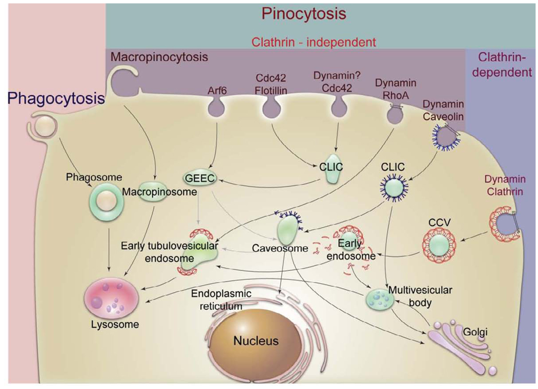
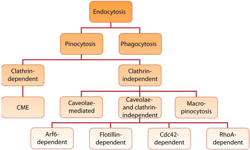
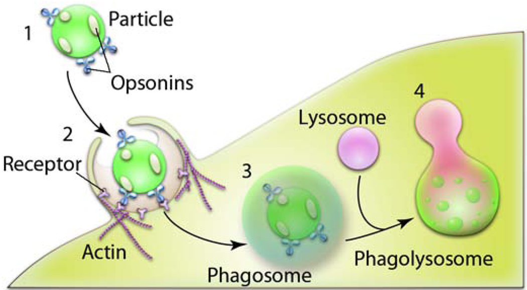
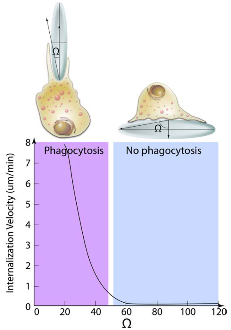
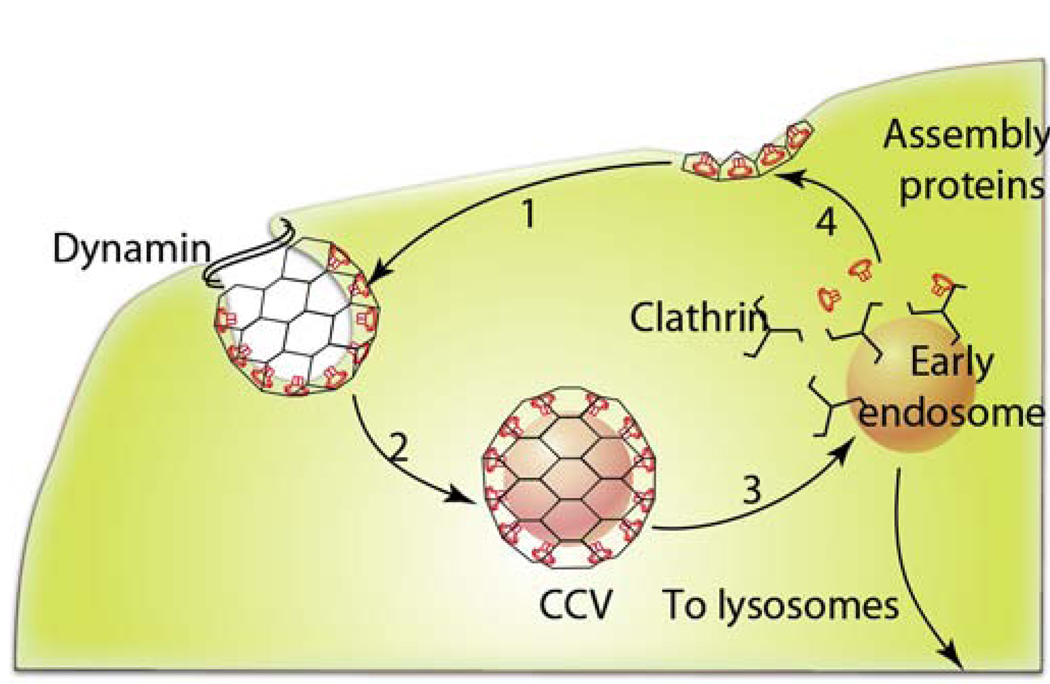
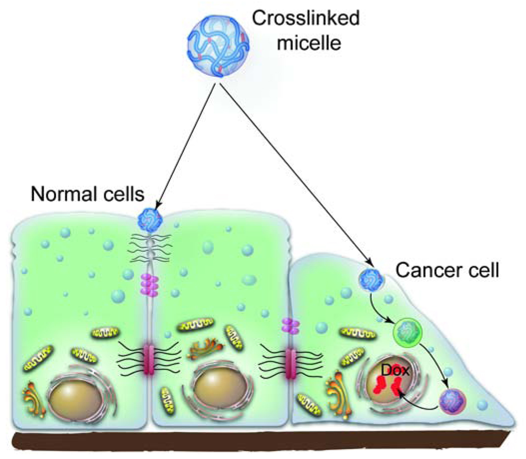
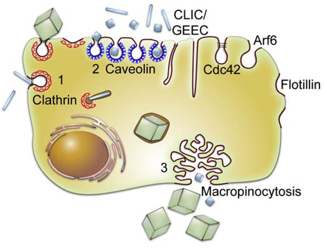
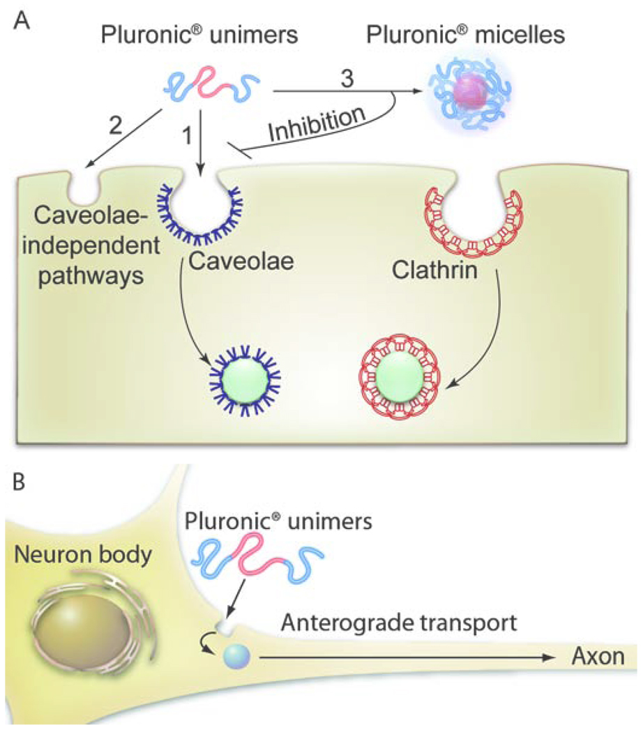
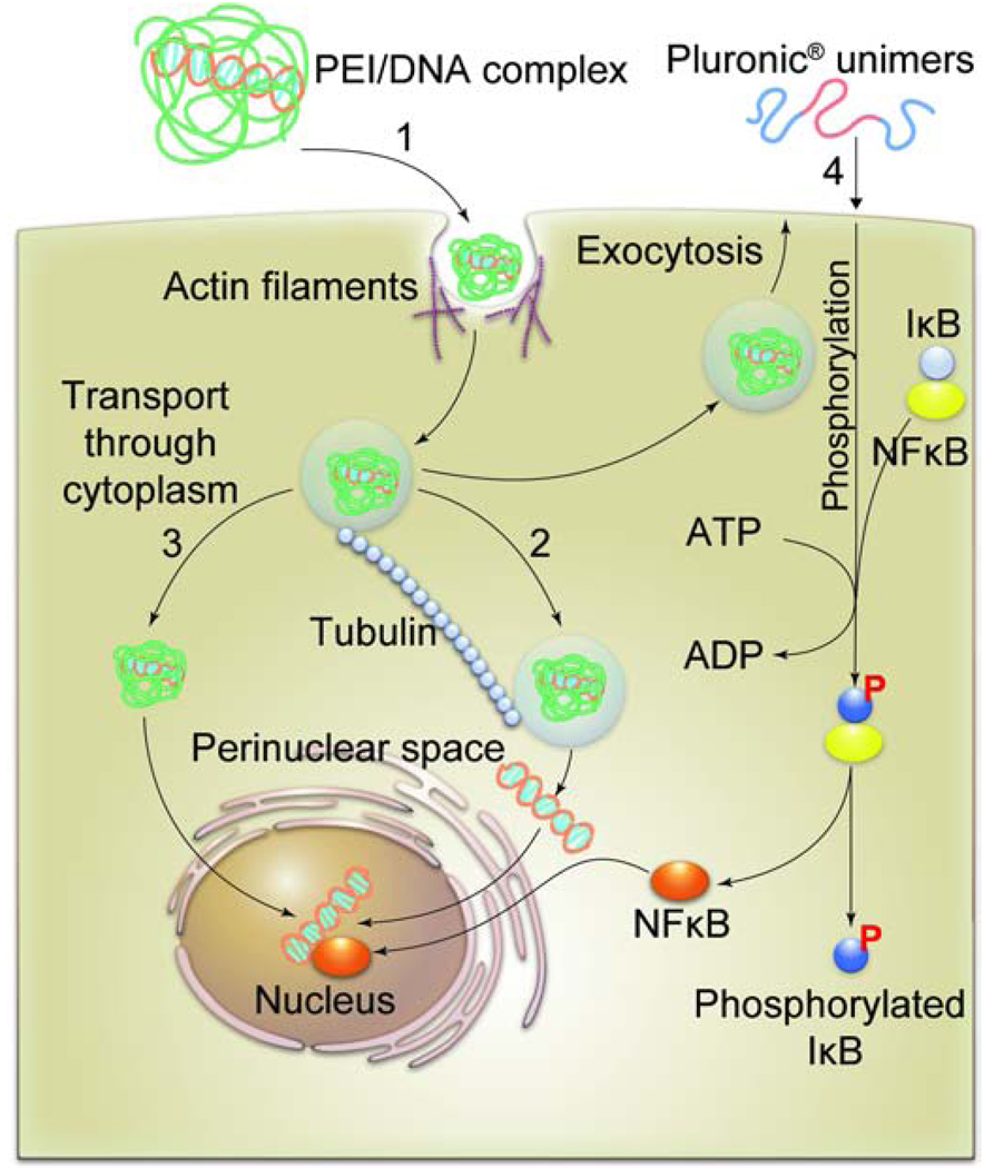
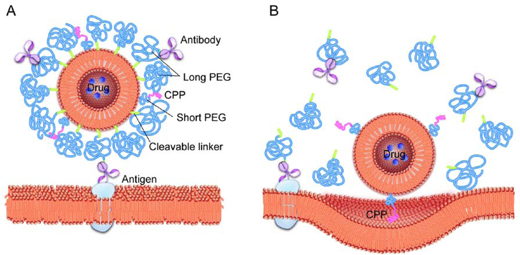
Novel nanomaterials are being developed to improve diagnosis and therapy of diseases through effective delivery of drugs, biopharmaceutical molecules and imaging agents to target cells in disease sites. Such diagnostic and therapeutic nanomaterials, also termed "nanomedicines", often require site-specific cellular entry to deliver their payload to sub-cellular locations hidden beneath cell membranes. Nanomedicines can employ multiple pathways for cellular entry, which are currently insufficiently understood. This review, first, classifies various mechanisms of endocytosis available to nanomedicines including phagocytosis and pinocytosis through clathrin-dependent and clathrin-independent pathways. Second, it describes the current experimental tools to study endocytosis of nanomedicines. Third, it provides specific examples from recent literature and our own work on endocytosis of nanomedicines. Finally, these examples are used to ascertain 1) the role of particle size, shape, material composition, surface chemistry and/or charge for utilization of a selected pathway(s); 2) the effect of cell type on the processing of nanomedicines; and 3) the effect of nanomaterial-cell interactions on the processes of endocytosis, the fate of the nanomedicines and the resulting cellular responses. This review will be useful to a diverse audience of students and scientists who are interested in understanding endocytosis of nanomedicines.


Figures

Fig. 1
Fig. 1. Different mechanisms of endocytosis
There are multiple pathways for cellular entry of particles and solutes. The picture of endocytosis trafficking is actively researched and evolving [11], [12]. In all cases the initial stage of endocytosis proceeds from the plasma membrane portals of cellular entry and involves engulfment of cargo into intracellular vesicles. The second stage often involves sorting of the cargo through endosomes. It is followed by the final stage during which the cargo is delivered to its final destination, recycled to extracellular milieu or delivered across cells (not shown). The figure is a simplified representation of complex trafficking mechanisms and their cross-talks. More details for stages of phagocytosis and CME are presented in Fig. 3 and Fig. 5. Abbreviations are: CCV, clathrin coated vesicles, CLIC, clathrin-independent carriers; GEEC, GPI-anchored protein-enriched compartment; GPI, glycophosphatidylinositol, MVB, multivesicular body.
Fig. 2
Fig. 2
Classification of endocytosis based on endocytosis proteins that are involved in the initial entry of particles and solutes.
Fig. 3
Fig. 3. Stages of phagocytosis of particles
1) Particles undergo recognition in the bloodstream through opsonization i.e. adsorption of proteins (immunoglubulins (Ig) G (and M), complement components (C3, C4, C5); blood serum proteins (including laminin, fibronectin, etc.). 2) Opsonized particles attach onto the cell membrane through receptors present on the cell surface of a phagocyte. 3) The particles are ingested into phagosomes. 4) The phagosomes mature, fuse with lysosomes and become acidified, leading to the enzyme-rich phagolysosomes where the particles are prone to degradation.
Fig. 4
Fig. 4. Effect of particle geometry on phagocytosis
The entry of a nanoparticles inside macrophages depends on the angle between the membrane normal at the point of initial contact and the line defining the particle curvature at this point (Ω). The internalization velocity is positive at Ω ≤ 45°, which indicates that the particle undergoes internalization. As the angle exceeds critical value ≈ (45°) the internalization velocity is zero, the macrophages lose the ability to entrap particles and start spreading over the particle.
Fig. 5
Fig. 5. Schematic representation of CME
1) The assembly proteins, AP-2 and AP180 are targeted to the plasma membrane where they mediate clathrin assembly. Upon that clathrin triskelions polymerize into a polygonal lattice, which helps to deform the plasma membrane into a coated pit. 2) Dynamin, a multidomain GTPase, is recruited to the necks of coated pits, where it assembles into a spiral collar. Upon hydrolysis of GTP dynamic collar promotes scission of the membrane and release of the vesicle known as CCV. 3) The next step involves uncoating of CCV and formation of an early endosomes, which are then routed towards the lysosomes as shown in Fig. 1. 4) The coat constituents are recycled for reuse.
Fig. 6
Fig. 6. Pathways of intracellular trafficking of cl-micelles in normal and cancer epithelial cells
The cl-micelles carrying a drug, doxorubucin (Dox), in normal epithelial cells were shown to sequester at the apical surface of the cell membrane near the TJs. However, during cancer progression the TJs are lost. As a result the cancer epithelial cells internalize the cl-micelles through caveolae. The cl-micelles are then routed to the lysosomes where the drug is released through a pH-dependent mechanism. The released drug accumulates in the nucleus and kills the cancer cells.
Fig. 7
Fig. 7. Cellular entry of PRINT nano- and microparticles
PRINT nanoparticles of all shapes utilize multiple pathways to gain cellular entry including CME (1), caveolae-mediated endocytosis (2) and, to a lesser extent, macropinocytosis (3). The shape of the particles appears to be important in regulating the rate of their cellular entry (not shown). Cube-shaped PRINT microparticles also utilize multiple routes of cellular entry but their macropinocytosis appears to be the most prominent.
Fig. 8
Fig. 8. The entry mechanisms of Pluronic® block copolymers in (A) epithelial cells and (B) neurons
A. In cells displaying the caveolae pathway Pluronic® P85 unimers enter through caveolae-mediated endocytosis (1). In cells devoid of caveolae, such as confluent MDCK cells, the block copolymer unimers can also enter through caveolae-independent pathways (2). Once the concentration of the block copolymer increases above the CMC the micelles are formed, which enter through the CME (3). Under these conditions the block copolymer inhibits the caveolae-mediated endocytosis. B. In primary neurons (also devoid of the caveolae) the Pluronic® P85 unimers enter the cell body from where they undergo anterograde trafficking to the axons/dendrites.
Fig. 9
Fig. 9. DNA delivery to the nucleus
1). PEI/DNA complexes internalize into cells utilizing actin-dependent pathway. 2). The endosomes containing PEI/DNA complexes travel inside the cytoplasm along the microtubules and reach the perinuclear space where the DNA is released through an unknown mechanism. 3). An alternative mechanism may involve direct release of PEI/DNA complex from endosomal/lysosomal compartments, followed by the transport of the complex through the cytoplasm and to the nucleus. 4). The DNA import to the nucleus can be enhanced by activating cellular signaling by Pluronic® block copolymers. In this case Pluronics® bind with the cell membranes and activate phosphorylation of IκB by an IκB-kinase (not shown). The phosphorylated IκB dissociates from its complex with NFκB. The released active NFκB enhances transport of DNA into nucleus in a promoter-dependent fashion.
Fig. 10
Fig. 10. Targeting of stimuli-sensitive double-targeted liposome to tumors
A. The surface of the drug-loaded liposome is modified with a cell-penetrating peptide (CPP) attached via relatively short PEG chains. This peptide is masked by long PEG chains anchored to the liposome surface via. pH-sensitive cleavable links. Some of the long PEG chains are decorated with the antibody specific to the tumor antigen. The antibody is exposed and can bind with the antigen at the tumor cell surface. B. Inside the acidic microenvironment of the tumor the long PEG chains and the antibody conjugates are detached from the liposome resulting in exposure of the CPP. The CPP interacts with the cell membrane and facilitates endocytosis of the drug-loaded liposomes into tumor cells.

Similar articles

Cited by

References

    1. Kabanov AV, Alakhov VY. Pluronic (R) block copolymers in drug delivery: From micellar nanocontainers to biological response modifiers. Critical Reviews in Therapeutic Drug Carrier Systems. 2002;19:1–72. - PubMed
    1. Duncan R. The dawning era of polymer therapeutics. Nature Review Drug Discovery. 2003;2:347–360. - PubMed
    1. Farokhzad OC, Langer R. Nanomedicine: Developing smarter therapeutic and diagnostic modalities. Advanced Drug Delivery Reviews. 2006;58:1456–1459. - PubMed
    1. Davis ME, Chen Z, Shin DM. Nanoparticle therapeutics: an emerging treatment modality for cancer. Nature Review Drug Discovery. 2008;7:771–782. - PubMed
    1. Peer D, Karp JM, Hong S, Farokhzad OC, Margalit R, Langer R. Nanocarriers as an emerging platform for cancer therapy. Nature Nanotechnology. 2007;2:751–760. - PubMed

Publication types