. 2010 Mar 16;17(3):235-48.
doi: 10.1016/j.ccr.2009.12.046.

DNA polymerases as potential therapeutic targets for cancers deficient in the DNA mismatch repair proteins MSH2 or MLH1

Affiliations

DNA polymerases as potential therapeutic targets for cancers deficient in the DNA mismatch repair proteins MSH2 or MLH1

Sarah A Martin et al. Cancer Cell. .

Abstract

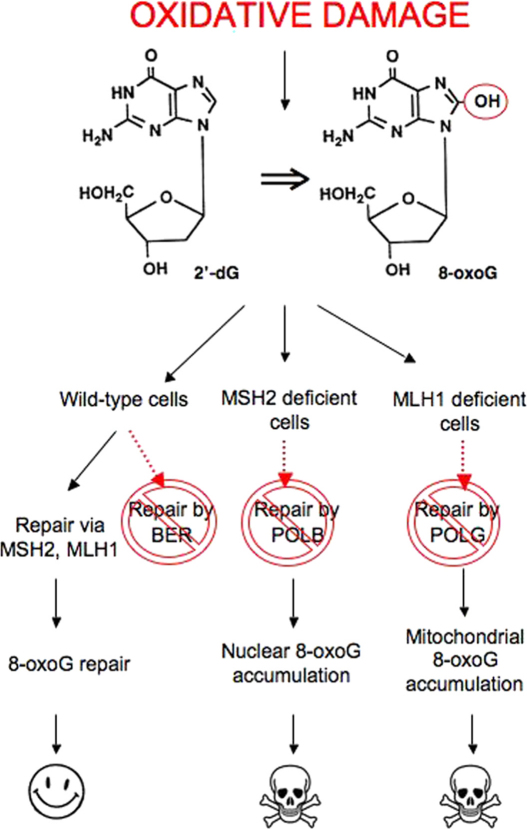
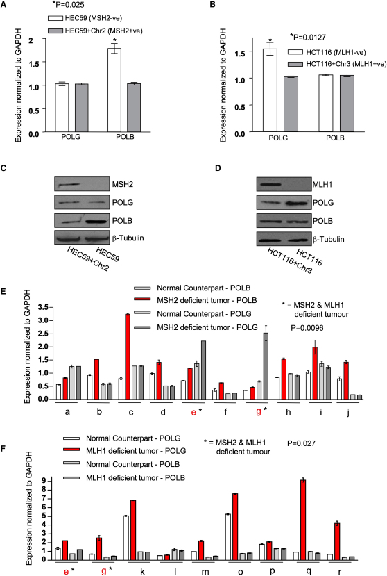
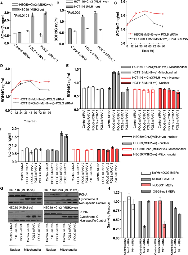
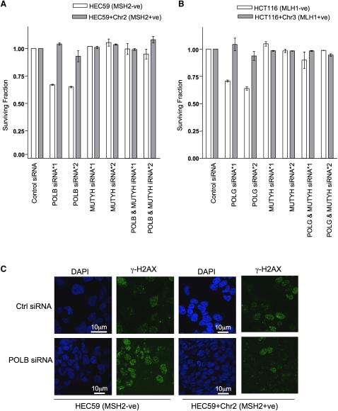
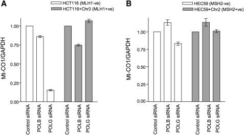
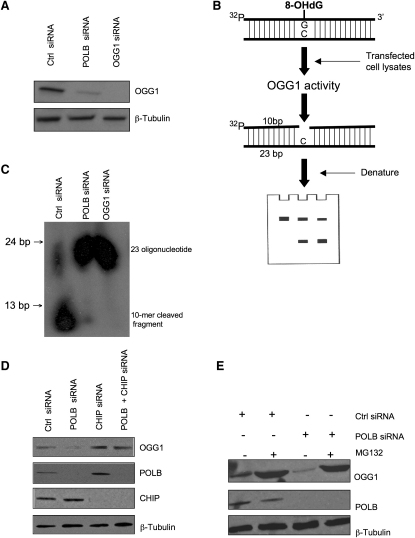
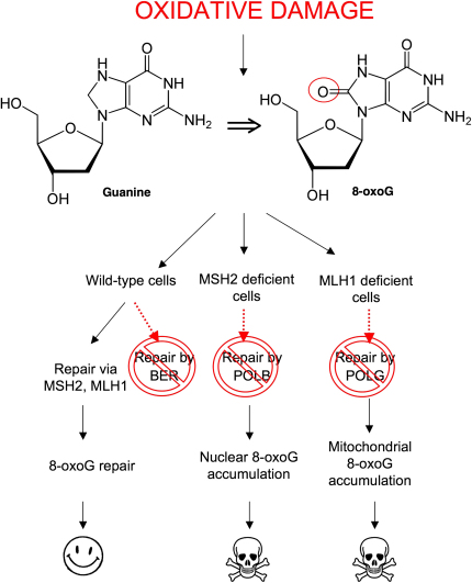
Synthetic sickness/lethality (SSL) can be exploited to develop therapeutic strategies for cancer. Deficiencies in the tumor suppressor proteins MLH1 and MSH2 have been implicated in cancer. Here we demonstrate that deficiency in MSH2 is SSL with inhibition of the DNA polymerase POLB, whereas deficiency in MLH1 is SSL with DNA polymerase POLG inhibition. Both SSLs led to the accumulation of 8-oxoG oxidative DNA lesions. MSH2/POLB SSL caused nuclear 8-oxoG accumulation, whereas MLH1/POLG SSL led to a rise in mitochondrial 8-oxoG levels. Both SSLs were rescued by silencing the adenine glycosylase MUTYH, suggesting that lethality could be caused by the formation of lethal DNA breaks upon 8-oxoG accumulation. These data suggest targeted, mechanism-based therapeutic approaches.


Figures

None
Graphical abstract
Figure 1
Figure 1
MSH2 and MLH1 Deficiencies Are Synthetically Sick/Lethal with Silencing of DNA Polymerases (A) Cell lysates from HCT116 and HCT116+Chr3 cells were analyzed by western blotting using MLH1- and β-tubulin-specific antibodies. (B) Cell lysates from HEC59 and HEC59+Chr2 cells were analyzed by western blotting using MSH2- and β-tubulin-specific antibodies. (C) HCT116 (MLH1-deficient) and HCT116+Chr3 (MLH1-proficient) cells were transfected with siRNAs targeting DNA polymerases, and cell viability was estimated 5 days later using CellTiter-Glo reagent. (D) HEC59 and HEC59+Chr2 cells were transfected with siRNAs targeting DNA polymerases, and cell viability was estimated 5 days later using CellTiter-Glo reagent. (E) HEC59 cells were transfected with siRNA, and cell lysates were analyzed by western blotting 72 hr later. POLB- and β-tubulin-specific antibodies were used as shown. (F) HCT116 cells were transfected with siRNA, and cell lysates were analyzed by western blotting 72 hr later. POLG and β-tubulin antibodies were used as shown. (G) HEC59 and HEC59+Chr2 cells were transfected with either control siRNA or siRNA targeting POLB, and clonogenic assays were performed. (H) HCT116 and HCT116+Chr3 cells were transfected with either control siRNA or siRNA targeting POLG, and clonogenic assays were performed. Error bars for each individual experiment represent standard errors of the mean. See also Figure S1.
Figure 2
Figure 2
MSH2 and MLH1 Deficiencies Are Associated with Particular Increases in DNA Polymerase Expression (A) POLB mRNA levels were analyzed by qRT-PCR using GAPDH expression as a control. p = 0.0254 compared to the MSH2-proficient HEC59+Chr2 cells (Student's t test). (B) POLG mRNA levels were analyzed by qRT-PCR using GAPDH expression as a control. p = 0.0127 compared to the MLH1-proficient HCT116+Chr3 cells (Student's t test). (C) Cell lysates from HEC59 and HEC59+Chr2 cells were analyzed by western blotting using MSH2-, POLB-, POLG-, and β-tubulin-specific antibodies as shown. (D) Cell lysates from HCT116 and HCT116+Chr3 cells were analyzed by western blotting using MLH1-, POLG-, POLB-, and β-tubulin-specific antibodies as shown. (E and F) Polymerase mRNA expression in MSH2- and MLH1-deficient tumors and adjacent nontumor material from the same patient was determined by qRT-PCR. MSH2-deficient tumors are shown in (E) and MLH1-deficient tumors are shown in (F). Patient samples are labeled alphabetically, with patients e and g having tumors with both MSH2 and MLH1 deficiency (labeled by ). MSH2 and MLH1 protein expression was determined by immunohistochemical analysis. The relative polymerase mRNA levels were measured using qRT-PCR and normalized to a housekeeping gene, GAPDH. (E) POLB expression was elevated in MSH2-deficient tumors, compared to patient-matched nontumor material; p = 0.0096 (pairwise Student's t test). (F) POLG expression was elevated in MLH1-deficient tumors, compared to patient-matched nontumor material; p = 0.027 (pairwise Student's t test). Error bars for each individual experiment represent standard errors of the mean. See also Figure S2.
Figure 3
Figure 3
Increased 8-OxoG Accumulation Correlates with Polymerase Inhibition and MSH2 or MLH1 Deficiency (A) HEC59 and HEC59+Chr2 cells were transfected with siRNA. Seventy-two hours after transfection, DNA was isolated from cells and analyzed for 8-oxoG accumulation using an ELISA assay. Oxidized lesions were quantified according to a standard curve generated using known amounts of 8-oxoG. Assays were performed in triplicate. p ≤ 0.0101 compared to the similarly transfected MSH2-proficient HEC59+Chr2 cells (Student's t test). (B) HCT116 and HCT116+Chr3 cells were transfected with siRNA and analyzed as in (A). p ≤ 0.002 compared to the similarly transfected MLH1-proficient HCT116+Chr3 cells (Student's t test). (C) HEC59 and HEC59+Chr2 cells were transfected with siRNA and DNA and analyzed as in (A) over a 96 hr time course. Assays were performed in triplicate. (D) HCT116 and HCT116+Chr3 cells were transfected with siRNA and DNA and analyzed as in (B) over a 96 hr time course. Assays were performed in triplicate. (E) HCT116 and HCT116+Chr3 cells were transfected with siRNA. Nuclear and mitochondrial DNA isolated from transfected cells were analyzed for 8-oxoG accumulation as in (B). Assays were performed in triplicate. (F) HEC59 and HEC59+Chr2 cells were transfected with siRNA. Nuclear and mitochondrial DNA isolated from transfected cells were analyzed for 8-oxoG accumulation as in (A). Assays were performed in triplicate. (G) Validation of nuclear and mitochondrial fractionation. Cells were transfected with siRNA, and nuclear and mitochondrial lysates were analyzed by western blotting. PCNA- and cytochrome c-specific antibodies were used to determine nuclear and mitochondrial fractionations, respectively. (H) Silencing of Mlh1 is synthetically lethal with loss of a mitochondrial isoform of Ogg1, whereas silencing of Msh2 is synthetically lethal with loss of a nuclear isoform of Ogg1. Fibroblasts expressing either a nuclear isoform of Ogg1, a mitochondrial isoform of Ogg1, or both Ogg1 isoforms were transfected with either nontargeting control siRNA, Msh2 siRNA, or Mlh1 siRNA. After 5 days, cell survival was estimated using CellTiter-Glo reagent. Error bars for each individual experiment represent standard errors of the mean. See also Figure S3.
Figure 4
Figure 4
MLH1/POLG and MSH2/POLB SSL Phenotypes Are Rescued by MUTYH Silencing (A) HEC59 and HEC59+Chr2 cells were transfected with either control, POLB, MUTYH siRNA, or with siRNAs in combination, and clonogenic assays were performed. (B) HCT116 and HCT116+Chr3 cells were transfected with either control, POLB, MUTYH siRNA, or with siRNAs in combination, and clonogenic assays were performed. (C) HEC59 and HEC59+Chr2 cells were transfected with either control or POLB siRNA. Seventy-two hours after transfection, γ-H2AX foci were quantified by immunofluorescence. Nuclei are shown in blue and γ-H2AX foci are in green. The scale bars represent 10 μm. Error bars for each individual experiment represent standard errors of the mean. See also Figure S4.
Figure 5
Figure 5
Depletion of Mt-CO1 DNA upon Inhibition of POLG in MLH1-Deficient Cells (A) HCT116 and HCT116+Chr3 cells were transfected with either control, POLB, or POLG siRNA. Mt-CO1 DNA levels were analyzed by qPCR with GAPDH used as a control. (B) HEC59 and HEC59+Chr2 cells were transfected with either control, POLB, or POLG siRNA. Mt-CO1 DNA levels were analyzed by qPCR with GAPDH used as a control. Error bars for each individual experiment represent standard errors of the mean.
Figure 6
Figure 6
OGG1 Cleavage Activity Is Decreased in the Absence of POLB Expression (A) HeLa cells were transfected with siRNA, and lysates were analyzed for OGG1 expression 72 hr later. (B) Schematic model for in vitro OGG1 assay. Briefly, protein was isolated from transfected cells. The DNA substrate is a 23 base oligonucleotide containing 8-oxoG at its 11th base, labeled with 32P at its 5′ end, and annealed to its complementary strand (containing dC at the opposite base position to the 8-oxoG). Upon cleavage of the substrate by the OGG1 enzyme, the oligonucleotides were electrophoresed on a denaturing PAGE gel, followed by autoradiography. (C) HeLa cells were transfected with either control, POLB, or OGG1 siRNA and incubated with an oligonucleotide substrate containing 8-oxoG, as described above. The oligonucleotides were electrophoresed and a 10 base fragment (labeled cleavage product) was revealed in addition to the original 23 base oligonucleotide. Autoradiography revealed that in the absence of POLB expression, cleavage of the 8-OHdG lesion was significantly decreased. (D) HeLa cells were transfected with siRNA, and cell lysates were analyzed 72 hr later. OGG1 expression was suppressed by POLB siRNA but rescued by combined silencing of POLB and CHIP. (E) Cell lysates from HeLa cells were transfected with siRNA and, after 48 hr, cells were treated with MG132 (50 μM). Lysates were analyzed 18 hr later by western blotting. Antibodies directed against OGG1, POLB, and β-tubulin were used to demonstrate reduction in expression of OGG1 after transfection with POLB siRNA, which was rescued by treatment with the proteasomal inhibitor MG132. See also Figure S5.
Figure 7
Figure 7
A Model for the Selective Effects of DNA Polymerase Inhibition in MLH1- or MSH2-Deficient Cells Oxidized DNA lesions, including 8-oxoG, can be repaired by either MSH2/MLH1-dependent processes or BER. In wild-type cells, inhibition of POLB or POLG leads to repair of these lesions by MSH2/MLH1. In the absence of MSH2, POLB is essential for 8-oxoG repair. Inhibition of POLB in MSH2-deficient cells leads to the accumulation of 8-oxoG in nuclear DNA. Cells harboring these unrepaired lesions may permanently arrest or die. These effects may be mediated by MUTYH, which in attempting to reverse C→A transversions opposite oxidized bases ultimately causes the formation of single-strand DNA breaks which in turn cause the formation of lethal double-strand DNA breaks. In cells with MLH1 deficiency, POLG inhibition leads to the accumulation of 8-oxoG in mitochondrial DNA. Again, this accumulation either becomes incompatible with viability or limits the cell's replicative potential.

References

    1. Aquilina G., Ceccotti S., Martinelli S., Hampson R., Bignami M. N-(2-chloroethyl)-N′-cyclohexyl-N-nitrosourea sensitivity in mismatch repair-defective human cells. Cancer Res. 1998;58:135–141. - PubMed
    1. Argueso J.L., Smith D., Yi J., Waase M., Sarin S., Alani E. Analysis of conditional mutations in the Saccharomyces cerevisiae MLH1 gene in mismatch repair and in meiotic crossing over. Genetics. 2002;160:909–921. - PMC - PubMed
    1. Arnold C.N., Goel A., Boland C.R. Role of hMLH1 promoter hypermethylation in drug resistance to 5-fluorouracil in colorectal cancer cell lines. Int. J. Cancer. 2003;106:66–73. - PubMed
    1. Bettstetter M., Dechant S., Ruemmele P., Grabowski M., Keller G., Holinski-Feder E., Hartmann A., Hofstaedter F., Dietmaier W. Distinction of hereditary nonpolyposis colorectal cancer and sporadic microsatellite-unstable colorectal cancer through quantification of MLH1 methylation by real-time PCR. Clin. Cancer Res. 2007;13:3221–3228. - PubMed
    1. Boiteux S., Radicella J.P. The human OGG1 gene: structure, functions, and its implication in the process of carcinogenesis. Arch. Biochem. Biophys. 2000;377:1–8. - PubMed

Publication types