. 2010 Mar 12;37(5):656-67.
doi: 10.1016/j.molcel.2010.02.012.

Topological layers in the HIV-1 gp120 inner domain regulate gp41 interaction and CD4-triggered conformational transitions

Affiliations

Topological layers in the HIV-1 gp120 inner domain regulate gp41 interaction and CD4-triggered conformational transitions

Andrés Finzi et al. Mol Cell. .

Abstract

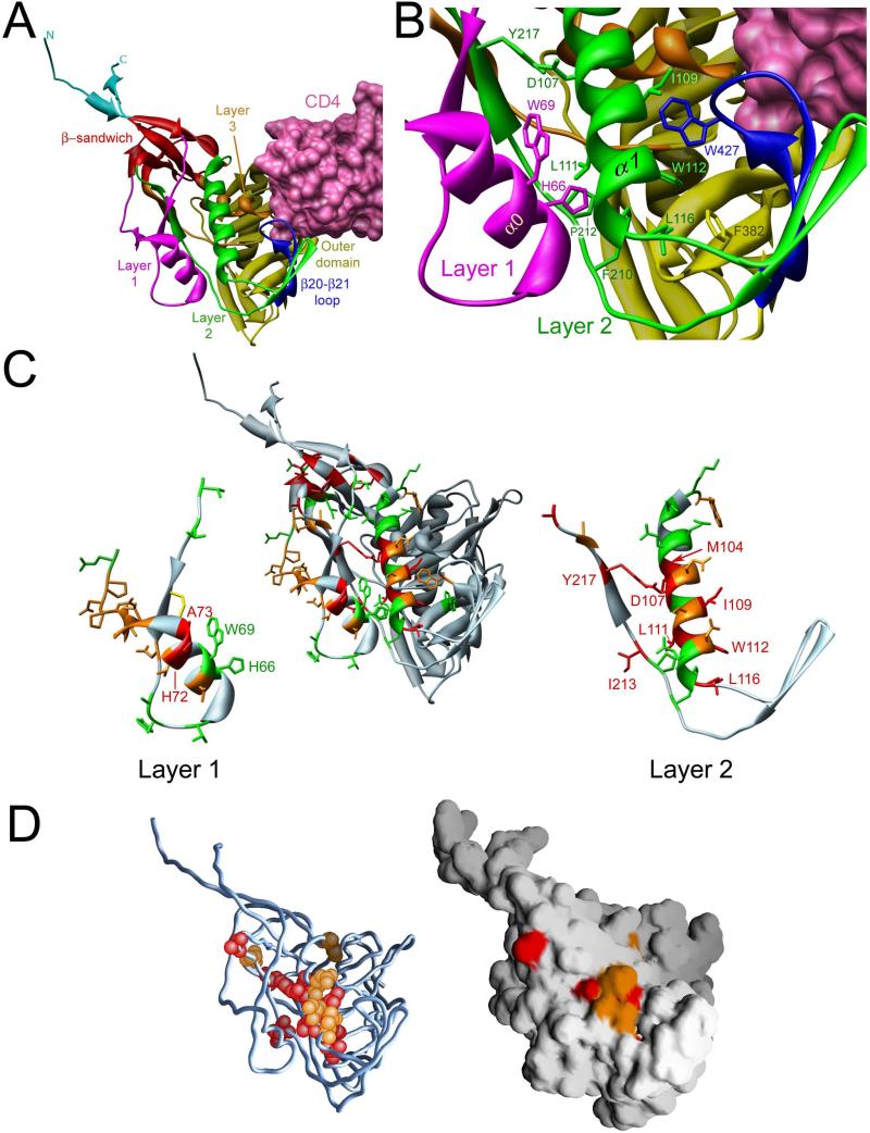
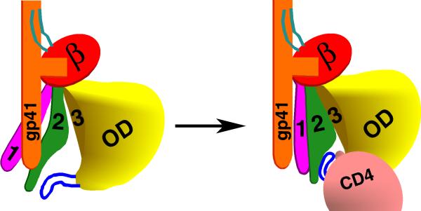
The entry of human immunodeficiency virus (HIV-1) into cells is initiated by binding of the gp120 exterior envelope glycoprotein to the receptor, CD4. How does CD4 binding trigger conformational changes in gp120 that allow the gp41 transmembrane envelope glycoprotein to mediate viral-cell membrane fusion? The transition from the unliganded to the CD4-bound state is regulated by two potentially flexible topological layers (layers 1 and 2) in the gp120 inner domain. Both layers apparently contribute to the noncovalent association of unliganded gp120 with gp41. After CD4 makes initial contact with the gp120 outer domain, layer 1-layer 2 interactions strengthen gp120-CD4 binding by reducing the off rate. Layer 1-layer 2 interactions also destabilize the activated state induced on HIV-1 by treatment with soluble CD4. Thus, despite lack of contact with CD4, the gp120 inner-domain layers govern CD4 triggering by participating in conformational transitions within gp120 and regulating the interaction with gp41.


Figures

Figure 1
Figure 1. Structure of the inner domain of HIV-1 gp120 in the CD4-bound conformation
A. The structure (Pancera et al., 2010) of HIV-1HXBc2 gp120 (ribbon) complexed with two-domain CD4 (pink molecular surface) is shown, from the approximate perspective of the Env gp trimer axis. The outer domain of gp120 is colored yellow. The N and C termini are colored cyan. The components of the gp120 inner domain are the β-sandwich (red) and three loop-like extensions: Layer 1 (magenta), Layer 2 (green) and Layer 3 (orange). The β20-β21 strands of gp120 (blue) project from the outer domain and, in the CD4-bound conformation, compose two of the strands of the four-stranded bridging sheet. The other two strands of the bridging sheet are derived from the distal portion of Layer 2. B. A close-up view of the interactions of Layer 2 (green) with Layer 1 (magenta), and with the β20-β21 loop (blue), is shown from the same perspective as in A. The surface of CD4 (pink) is visible in the upper right-hand corner of the image. C. The center image is a ribbon diagram of the CD4-bound HIV-1 gp120 glycoprotein, shown in the same orientation as that in A. The ribbon and side chain residues that were altered in this and a previous study (Yang et al., 2003) are colored according to the gp120-gp41 association index (Red: association index < 0.5; Orange: 0.5 ≤ association index < 0.7; Green: association index ≥ 0.7). In the left- and right-hand images, Layer 1 and Layer 2 are separated from the rest of the gp120 glycoprotein. The disulfide bond in Layer 1 between cysteines 54 and 74 is colored yellow. D. The gp120 glycoprotein, in the CD4-bound conformation, is shown from the perspective in A. In the left image, on the Cα trace of gp120, the atoms of Layer 2 residues with association indices less than 0.5 are colored red; residues with association indices between 0.5 and 0.7 are colored orange. The image at the right shows the solvent-accessible surface of gp120, colored similarly.
Figure 2
Figure 2. Binding of gp120 receptors
A, B. The effects of alterations in the Layer 1-Layer 2 interface (A) or in Layer 2 (B) on gp120 recognition by CD4-Ig were examined. Normalized amounts of radiolabeled wild-type mutant gp120 glycoproteins were incubated with increasing concentrations of CD4-Ig for 2 hours at 37°C. The precipitates were washed, run on SDS-polyacrylamide gels, and analyzed by densitometry. All values were normalized to a saturating value for the wt gp120. Representative results from at least two independent experiments are shown. C, D. Similar amounts of radiolabeled gp120 glycoproteins were incubated in the absence (C) or presence (D) of 200 nM sCD4 prior to addition to cells expressing CCR5. After two hours at 37°C, the amount of bound mutant gp120 was determined and normalized to the observed amount of bound wt gp120. Incubation with sCD4 increased the binding of wt gp120 8-fold. The data shown represent the means +/- SEM of two independent experiments.
Figure 3
Figure 3. Sensitivity of HIV-1 Env gp variants to soluble CD4 (sCD4)
A-C. Recombinant HIV-1 expressing luciferase and bearing wt or mutant HIV-1 Env gps were normalized by reverse transcriptase activity. Equal amounts of viruses were incubated with serial dilutions of sCD4 at 37°C for 1 hour prior to infection of Cf2Th-CD4/CCR5 cells. Infectivity at each dilution of sCD4 tested is shown as the percent of infection without sCD4 for each particular mutant. Duplicate samples were analyzed; data shown are representative of those obtained in at least three independent experiments. D, E. The sCD4-induced shedding of gp120 from HIV-1 Env gps expressed on the cell surface was measured. Transfected 293T cells were metabolically labeled with [35S]-methionine/cysteine for 16 hours in the presence of increasing concentrations of sCD4 (0 to 200 nM). Cell lysates were precipitated with a mixture of sera from HIV-1-infected individuals. Precipitates were analyzed by SDS-PAGE, autoradiography and densitometry. Data shown represent the means +/− SEM obtained from two independent experiments.
Figure 4
Figure 4. Effect of soluble CD4 on HIV-1 infection of CD4-negative, CCR5-expressing cells
Recombinant luciferase-expressing HIV-1 with the indicated HIV-1 Env gps were normalized by reverse transcriptase activity and incubated with serial dilutions of sCD4 at 37°C for 1 hour prior to infection of Cf2Th-CCR5 cells. The level of infection is reported as Relative Light Units (RLU). For each data point, the infection was performed in duplicate; data shown represent the means +/- SEM derived from two independent experiments.
Figure 5
Figure 5. A model of the activation of the HIV-1 Env gps triggered by binding to the CD4 receptor
One of the three subunits of the HIV-1 Env gp trimer is depicted, oriented so that the viral membrane is at the top of the figure. The gp41 ectodomain is colored orange, the gp120 N and C termini cyan, the β-sandwich red, Layer 1 magenta, Layer 2 green, Layer 3 yellow, outer domain (OD) yellow, and the β20-β21 loop blue. In the unliganded state (left figure), Layer 1 and Layer 2 assume conformations different from that observed in the CD4-bound state (Pancera et al., 2010), allowing both layers to interact with the gp41 ectodomain. CD4 binding results in the apposition of Layer 1 and Layer 2 (right figure). This rearrangement of the gp120 inner domain slows the off-rate of CD4 and allows the gp41 ectodomain to undergo additional conformational changes necessary for HIV-1 entry.

References

    1. Alkhatib G, Combadiere C, Broder CC, Feng Y, Kennedy PE, Murphy PM, Berger EA. CC CKR5: a RANTES, MIP-1alpha, MIP-1beta receptor as a fusion cofactor for macrophage-tropic HIV-1. Science. 1996;272:1955–1958. - PubMed
    1. Allan JS, Coligan JE, Barin F, McLane MF, Sodroski JG, Rosen CA, Haseltine WA, Lee TH, Essex M. Major glycoprotein antigens that induce antibodies in AIDS patients are encoded by HTLV-III. Science. 1985;228:1091–1094. - PubMed
    1. Allan JS, Strauss J, Buck DW. Enhancement of SIV infection with soluble receptor molecules. Science. 1990;247:1084–8. - PubMed
    1. Binley JM, Sanders RW, Clas B, Schuelke N, Master A, Guo Y, Kajumo F, Anselma DJ, Maddon PJ, Olson WC, et al. A recombinant human immunodeficiency virus type 1 envelope glycoprotein complex stabilized by an intermolecular disulfide bond between the gp120 and gp41 subunits is an antigenic mimic of the trimeric virion-associated structure. J. Virol. 2000;74:627–43. - PMC - PubMed
    1. Chan DC, Fass D, Berger JM, Kim PS. Core structure of gp41 from the HIV envelope glycoprotein. Cell. 1997;89:263–273. - PubMed

Publication types

MeSH terms