Review
. 2010 Jun 18;584(12):2618-25.
doi: 10.1016/j.febslet.2010.03.017. Epub 2010 Mar 15.

Circadian clock control of the cellular response to DNA damage

Affiliations
Review

Circadian clock control of the cellular response to DNA damage

Aziz Sancar et al. FEBS Lett. .

Abstract

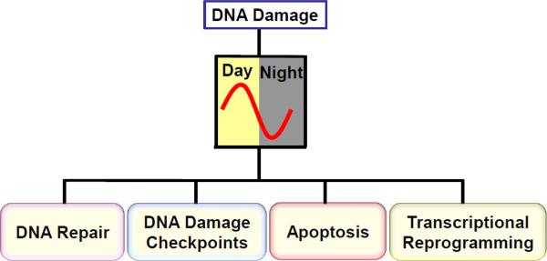
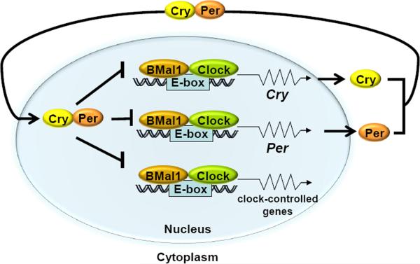
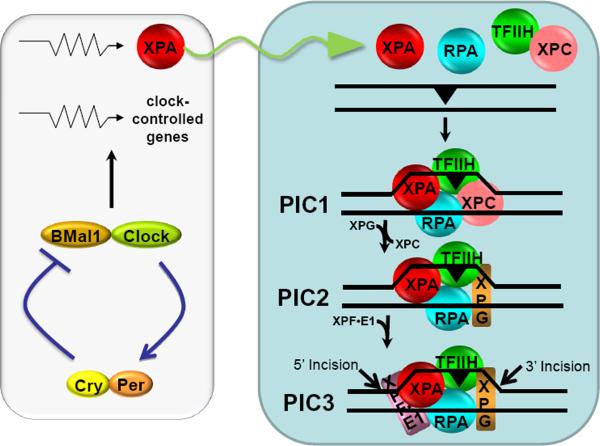
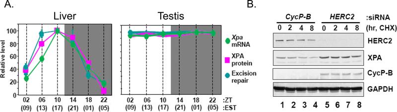
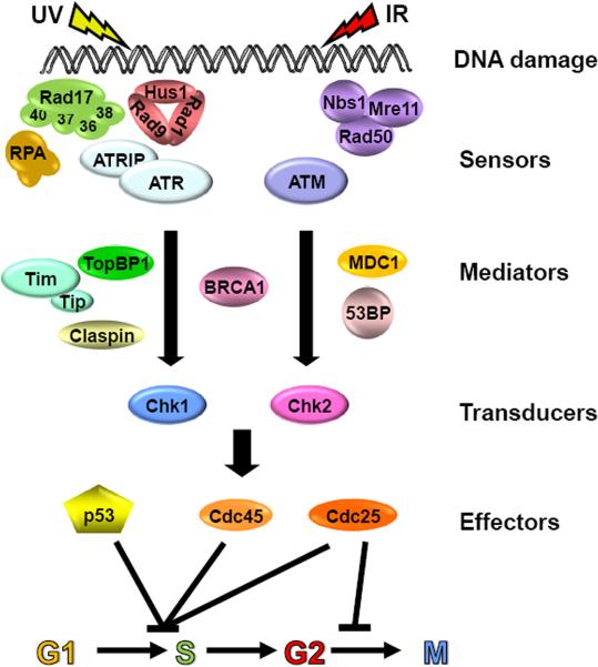
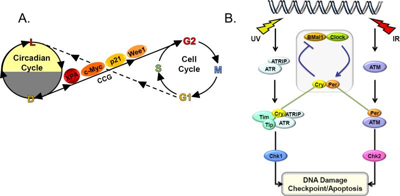
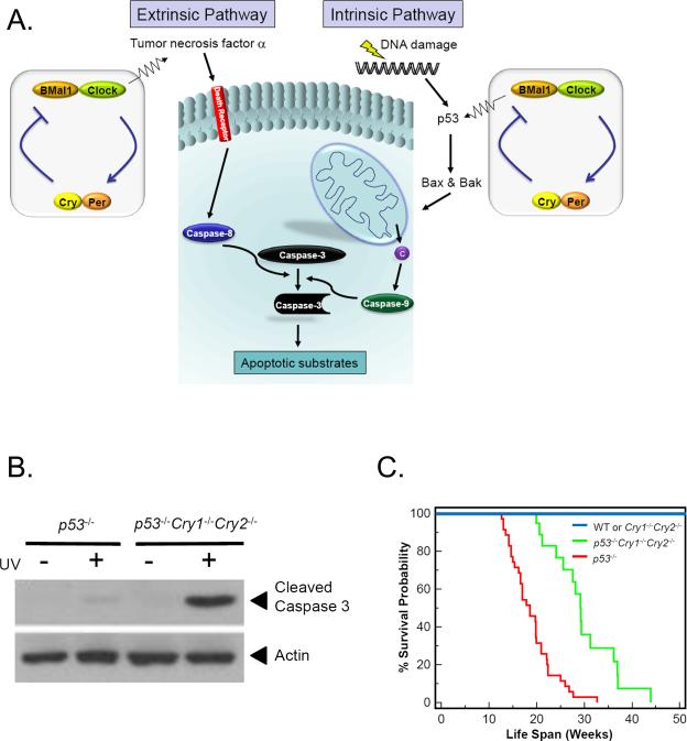
Mammalian cells possess a cell-autonomous molecular clock which controls the timing of many biochemical reactions and hence the cellular response to environmental stimuli including genotoxic stress. The clock consists of an autoregulatory transcription-translation feedback loop made up of four genes/proteins, BMal1, Clock, Cryptochrome, and Period. The circadian clock has an intrinsic period of about 24 h, and it dictates the rates of many biochemical reactions as a function of the time of the day. Recently, it has become apparent that the circadian clock plays an important role in determining the strengths of cellular responses to DNA damage including repair, checkpoints, and apoptosis. These new insights are expected to guide development of novel mechanism-based chemotherapeutic regimens.


Figures

Figure 1
Figure 1
Circadian gating of DNA damage responses. Major cellular response pathways to DNA damage including DNA repair, DNA damage checkpoints, apoptosis, and transcriptional reprogramming are gated by the circadian clock.
Figure 2
Figure 2
Mammalian molecular clock. The bHLH-PAS domain-containing proteins Clock and BMal1 make a heterodimer which bind to the E-boxes (CACGTG) in the promoters of the Per and Cry genes, as well as in the promoters of the clock controlled genes, such as the excision repair gene Xpa and the checkpoint gene Wee1, activating their transcription. The Cry and Per proteins dimerize and, after a time lag, enter the nucleus and inhibit Clock-BMal1-activated transcription of their own genes as well as of those of clock-controlled genes, thus generating an oscillatory pattern of gene expression. Modified from [67].
Figure 3
Figure 3
Model for nucleotide excision repair in humans and its regulation by the circadian clock. Excision of DNA damage is accomplished by sequential and partially overlapping functions of 6 core repair factors. Damage is recognized by cooperative activities of RPA, XPA, and XPC, followed by recruitment of TFIIH by XPC and XPA. The DNA around the damage site is unwound by the helicase activity of TFIIH to form a stable pre-incision complex 1 (PIC1) which recruits XPG, and XPC is displaced from the complex to form PIC2. Then XPF-ERCC1 is recruited to form PIC3. Within PIC3, XPG makes the 3′ incision 6±3 phosphodiester bonds 3′ and XPF makes the 5′ incision 20±5 phosphodiester bonds 5′ to the damage. The excised 24-32 nucleotide-long oligomer carrying the damage is released and the resulting gap is filled by DNA polymerases and ligated. The XPA protein which plays an essential role in damage recognition and is a rate-limiting factor is regulated by the clock, and as a consequence the daily oscillation of XPA (sinusoidal arrow) causes the entire excision repair activity to exhibit a daily rhythm, increasing during the day and decreasing during the night. Modified from [9, 66].
Figure 4
Figure 4
Circadian regulation of XPA and excision repair by the clock and the ubiquitin-proteasome system in the liver but not in testis. (A) Circadian rhythm of XPA transcript, protein, and nucleotide excision repair in mouse liver (left panel) but not testis (right panel) (ZT=0 is light on and ZT=12 is light off; EST=Eastern Standard Time) [23]. (B) HERC2 E3 ligase controls XPA stability. A549 cells were transfected with either cyclophilin-B siRNA (control) or HERC2 siRNA and then incubated with cycloheximide (CHX, a protein synthesis inhibitor) for the indicated times and the XPA protein levels were determined by immunoblotting [24].
Figure 5
Figure 5
DNA damage checkpoint pathways. The pathways encompass damage sensors, mediators, signal transducers, and effectors. DNA damage in the form of double-strand breaks induced by ionizing radiation or radiomimetic agents activates the ATM→Chk2 pathway. DNA damage by UV and UV-mimetic agents activates the ATR→Chk1 pathway. Modified from [6].
Figure 6
Figure 6
Two models for coupling the circadian clock to the cell cycle/checkpoint response. (A) Serial coupling. The XPA repair factor, the p21 and Wee1 cell cycle proteins, and the c-Myc transcription factor, which are involved in DNA repair, the cell cycle, and cellular proliferation, respectively, are encoded by clock-controlled genes (CCG), and hence DNA repair, checkpoint activation, cell cycle regulation, and cellular proliferation are gated by the clock (solid line). Conversely, the cell cycle influences the circadian cycle by halting transcription during mitosis, thus causing a phase shift of the circadian rhythm (broken line). (B) Direct coupling. The core circadian clock protein Cry, in conjunction with Tim participates in the ATR-Chk1 signaling pathway in response to UV and UV-mimetic agents. Similarly, Per1 participates in the ATM-Chk2 signaling pathway in response to IR and radiomimetic agents.
Figure 7
Figure 7
Regulation of apoptosis by the circadian clock. (A) The extrinsic and intrinsic apoptosis pathways. Circadian regulation of the extrinsic pathway occurs by regulating the synthesis of TNFα which binds to death receptors, which through death adaptors transduce the signal to caspase 8 which in turn activates executioner caspases including caspase 3. In the intrinsic pathway, DNA damage activates p53 (in a circadian regulated manner) which activates transcription of Bax and Bak pro-apoptotic members of Bcl2 family. Bax and Bak cause release of cytochrome c (c) from mitochondrial intermembrane space. Cytochrome c helps assemble Apaf1 into an apoptosome which leads to cleavage and activation of transducer caspase 9 which in turn cleaves and activates executioner caspase 3. (B) Elimination of Crys in immortal p53-deficient cells makes cells more sensitive to killing by apoptosis induced by genotoxic agents including UV [46]. (C) Cry mutations increase the life-span of p53-/- mice [46]. The Kaplan-Meier survival analysis of mice of the indicated genotype is shown. Significant differences were observed between p53–/– (n=35) and p53–/–Cry1–/–Cry2–/– (n=19) mice (P < 0.0001).

References

    1. Reppert SM, Weaver DR. Coordination of circadian timing in mammals. Nature. 2002;418:935–41. - PubMed
    1. Hastings MH, Reddy AB, Maywood ES. A clockwork web: circadian timing in brain and periphery, in health and disease. Nat Rev Neurosci. 2003;4:649–61. - PubMed
    1. Takahashi JS, Hong HK, Ko CH, McDearmon EL. The genetics of mammalian circadian order and disorder: implications for physiology and disease. Nat Rev Genet. 2008;9:764–75. - PMC - PubMed
    1. Sancar A. Regulation of the mammalian circadian clock by cryptochrome. J Biol Chem. 2004;279:34079–82. - PubMed
    1. Hughes M, Deharo L, Pulivarthy SR, Gu J, Hayes K, Panda S, Hogenesch JB. High-resolution time course analysis of gene expression from pituitary. Cold Spring Harb Symp Quant Biol. 2007;72:381–6. - PMC - PubMed

Substances