. 2010 Jul;121(1-2):88-97.
doi: 10.1016/j.jsbmb.2010.03.019. Epub 2010 Mar 20.

The nuclear vitamin D receptor controls the expression of genes encoding factors which feed the "Fountain of Youth" to mediate healthful aging

Affiliations

The nuclear vitamin D receptor controls the expression of genes encoding factors which feed the "Fountain of Youth" to mediate healthful aging

Mark R Haussler et al. J Steroid Biochem Mol Biol. 2010 Jul.

Abstract

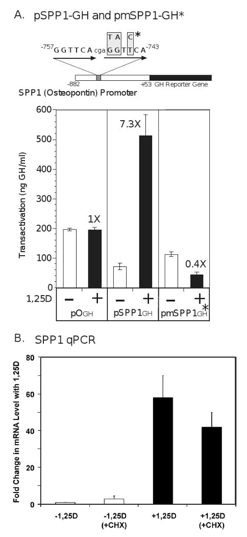
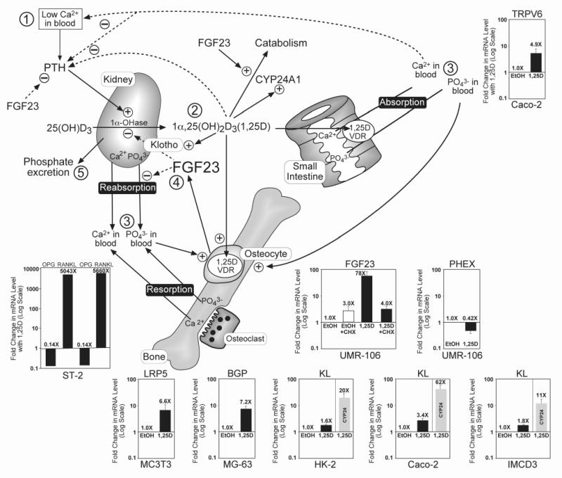
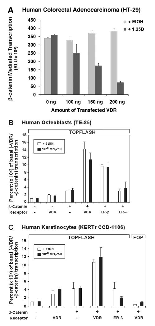
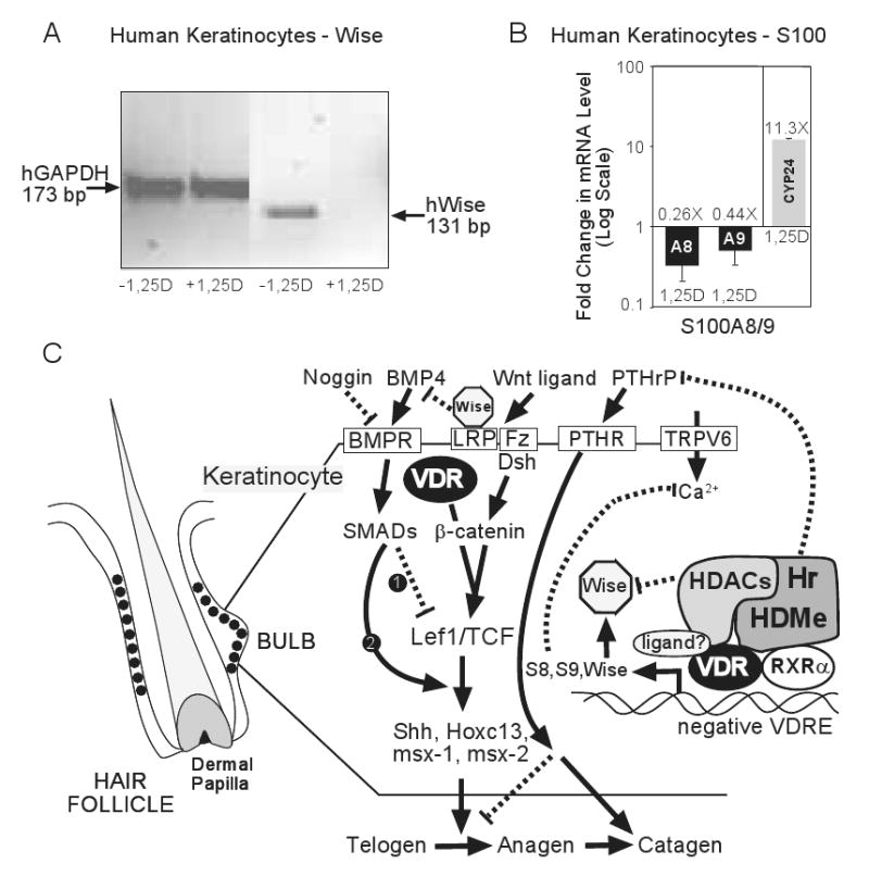
The nuclear vitamin D receptor (VDR) binds 1,25-dihydroxyvitamin D3 (1,25D), its high affinity renal endocrine ligand, to signal intestinal calcium and phosphate absorption plus bone remodeling, generating a mineralized skeleton free of rickets/osteomalacia with a reduced risk of osteoporotic fractures. 1,25D/VDR signaling regulates the expression of TRPV6, BGP, SPP1, LRP5, RANKL and OPG, while achieving feedback control of mineral ions to prevent age-related ectopic calcification by governing CYP24A1, PTH, FGF23, PHEX, and klotho transcription. Vitamin D also elicits numerous intracrine actions when circulating 25-hydroxyvitamin D3, the metabolite reflecting vitamin D status, is converted to 1,25D locally by extrarenal CYP27B1, and binds VDR to promote immunoregulation, antimicrobial defense, xenobiotic detoxification, anti-inflammatory/anticancer actions and cardiovascular benefits. VDR also affects Wnt signaling through direct interaction with beta-catenin, ligand-dependently blunting beta-catenin mediated transcription in colon cancer cells to attenuate growth, while potentiating beta-catenin signaling via VDR ligand-independent mechanisms in osteoblasts and keratinocytes to function osteogenically and as a pro-hair cycling receptor, respectively. Finally, VDR also drives the mammalian hair cycle in conjunction with the hairless corepressor by repressing SOSTDC1, S100A8/S100A9, and PTHrP. Hair provides a shield against UV-induced skin damage and cancer in terrestrial mammals, illuminating another function of VDR that facilitates healthful aging.


Figures

Fig. 1
Fig. 1
Osteopontin (SPP1) expression is induced by 1,25-VDR via a primary transcriptional mechanism involving a perfect direct repeat-3 (DR3) VDRE at -757 bp in the proximal promoter of the murine gene. (A) The mouse SPP1 proximal promoter obtained from Dr. David Dempster [94] was linked to the growth hormone (GH) reporter gene. ROS 17/2.8 rat osteosarcoma cells were cotransfected with an expression vector (1 μg) containing either the wild type version of the mouse osteopontin promoter (pSPP1GH) or its mutated VDRE version (pmSPP1GH*). The insertless vector p0GH was included as a control. Cells were treated at 12 hours post-transfection with either ethanol vehicle (-) or 1,25D at a concentration of 10 nM (+), and cell media assayed by RIA for growth hormone at 48 hours post-transfection. The data depicted represent the means (±SD) of two independent experiments with n=3 for each assay. The numbers at the top of the dark colored bars indicate the fold-induction of transcriptional activity following treatment with ligand. (B) UMR-106 rat osteosarcoma cells were treated with 100 nM 1,25D or ethanol vehicle for 24 hours in the presence (+CHX) or absence of 10 μM cycloheximide added 30 min prior to the 1,25D. All groups received both ethanol (for 1,25D) and DMSO (for cycloheximide) vehicles. RNA was isolated and reverse transcribed into cDNA that was assayed by real time PCR using custom primers against the coding region of SPP1, as described previously [16]. All values are normalized against the unregulated GAPDH control cDNA and are expressed as mean fold change ±SEM for three independent experiments, each performed in triplicate.
Fig. 2
Fig. 2
1,25D-VDR regulates the expression of many genes whose products effect bone mineral homeostasis and the integrity of the endoskeleton, as well as promote healthful aging. Insets show the induction or repression of each gene listed at the top, with the cell line denoted at the bottom of the inset. KL=klotho, a recognized longevity gene [51]. All mRNAs were quantitated via real time PCR as described previously [16], 24 hours after treatment of cells with 100 nM 1,25D or ethanol vehicle and expressed as fold-change (induction or repression) with 1,25D, plotted on a log scale with each value representing the mean ±SEM of at least three experiments each carried out in triplicate; an exception to the latter is ST2 mouse stromal cells, where RANKL and OPG mRNAs were measured in duplicate in the two experiments depicted in the inset and yielded essentially identical (0.14X) repression of OPG and over 5,000-fold induction of RANKL by 1,25D. Other cells utilized included Caco-2 human colon cancer, UMR-106 rat osteosarcoma, MG-63 human osteosarcoma, MC3T3 E1 mouse osteoblast-like, HK-2 human renal proximal tubule and mouse IMCD3 intermedullary collecting duct cells. The cycloheximide (CHX) protocol in the case of FGF23 regulation in UMR-106 cells was performed as described for SPP1 in Fig. 1B. Induction by 1,25D of LRP5, BGP and RANKL, plus repression of OPG, is discussed in the text as events that strengthen the skeleton via anabolic actions, extracellular matrix protein production and remodeling. The circled numbers in the schematic model portion of the figure represent the sequence of physiologic adjustments and feedback regulations of mineral metabolism after a reduction in blood calcium (➀) as follows: PTH-mediated enhancement of CYP27B1 (1α-OHase) and consequent rise in 1,25D hormone (➁); 1,25D-VDR action to cause calcium and phosphate absorption, reabsorption and resorption (➂) to raise both calcium and phosphate in blood; calcium and 1,25D negatively feedback (dashed lines) on PTH to close the calcemic loop, whereas 1,25D and phosphate cause osteocyte elaboration of FGF23 (➃), and 1,25D induces klotho to reduce 1,25D by inhibiting its production catalyzed by the 1α-OHase and inducing the CYP24A1 detoxification enzyme to accelerate its degradation; FGF23 also inhibits Npt2a/Npt2c-facilitated phosphate reabsorption to effect phosphate excretion (➄), protecting against hyperphosphatemia and ectopic calcification.
Fig. 3
Fig. 3
VDR modulates β-catenin signaling in a cell-specific manner to impact chemoprevention, bone remodeling, and hair cycling. (A) VDR transfection of human colorectal adenocarcinoma (HT-29) cells dose-responsively blunts β-catenin-mediated transcription in a 1,25D ligand-dependent fashion. β-catenin transcriptional activity was measured via TOPFLASH assay as described elsewhere [59], after treating human β-catenin-transfected HT-29 cells with 1 nM 1,25D for 24 hours. Error bars are ±SD, with n=3 for this representative experiment. VDR transfection of human osteoblast-like TE-85 (B), or human keratinocyte KERTr CCD-1106 (C) cells potentiates β-catenin transcriptional activity in the absence or presence of 10 nM 1,25D. TOPFLASH was assayed as described elsewhere [59], and FOP represents a loss of function mutant β-catenin responsive element construct that serves as a negative control.
Fig. 4
Fig. 4
VDR action in keratinocytes to sustain the mammalian hair cycle. (A) Repression of human Wise (SOSTDC1) gene expression by 1,25D in human keratinocytes (KERTr CCD-1106). Cultured cells were incubated with or without 10 nM 1,25D for 18 h. Levels of mRNA expression were evaluated by RT-PCR. Total RNA was isolated, reverse transcribed into cDNA, and the products analyzed via gel electrophoresis with primers designed to generate 131 bp and 173 bp respective coding region products from Wise and control GAPDH. (B) S100A8 (A8) and S100A9 (A9) genes are rapidly (3 h) repressed in KERTr CCD-1106 cells by 1,25D (10 nM) treatment in the face of an 11.3-fold induction of CYP24A1. RNA was isolated, reverse transcribed into cDNA, and analyzed by real time PCR using GAPDH as an uncontrolled normalizing factor. Error bars are ±SD with n=9. (C) Model for control of the mammalian hair cycle and the proposed role of VDR, RXRα, and Hr. See text for an explanation of the model.

Similar articles

Cited by

References

    1. Whitfield GK, Jurutka PW, Haussler CA, Hsieh JC, Barthel TK, Jacobs ET, Encinas Dominguez C, Thatcher ML, Haussler MR. Nuclear vitamin D receptor: structure-function, molecular control of gene transcription, and novel bioactions. In: Feldman D, Pike JW, Glorieux FH, editors. Vitamin D. Elsevier Academic Press; Oxford, UK: 2005. pp. 219–261.
    1. Whitfield GK, Dang HTL, Schluter SF, Bernstein RM, Bunag T, Manzon LA, Hsieh G, Dominguez CE, Youson JH, Haussler MR, Marchalonis JJ. Cloning of a functional vitamin D receptor from the lamprey (Petromyzon marinus), an ancient vertebrate lacking a calcified skeleton and teeth. Endocrinology. 2003;144(6):2704–2716. - PubMed
    1. Nehring JA, Zierold C, DeLuca HF. Lithocholic acid can carry out in vivo functions of vitamin D. Proc Natl Acad Sci U S A. 2007;104(24):10006–10009. - PMC - PubMed
    1. Makishima M, Lu TT, Xie W, Whitfield GK, Domoto H, Evans RM, Haussler MR, Mangelsdorf DJ. Vitamin D receptor as an intestinal bile acid sensor. Science. 2002;296(5571):1313–1316. - PubMed
    1. Echchgadda I, Song CS, Roy AK, Chatterjee B. Dehydroepiandrosterone sulfotransferase is a target for transcriptional induction by the vitamin D receptor. Mol Pharmacol. 2004;65(3):720–729. - PubMed

Publication types