. 2010 Mar 18;7(3):210-20.
doi: 10.1016/j.chom.2010.02.006.

Mycobacterium tuberculosis MycP1 protease plays a dual role in regulation of ESX-1 secretion and virulence

Affiliations

Mycobacterium tuberculosis MycP1 protease plays a dual role in regulation of ESX-1 secretion and virulence

Yamini M Ohol et al. Cell Host Microbe. .

Abstract

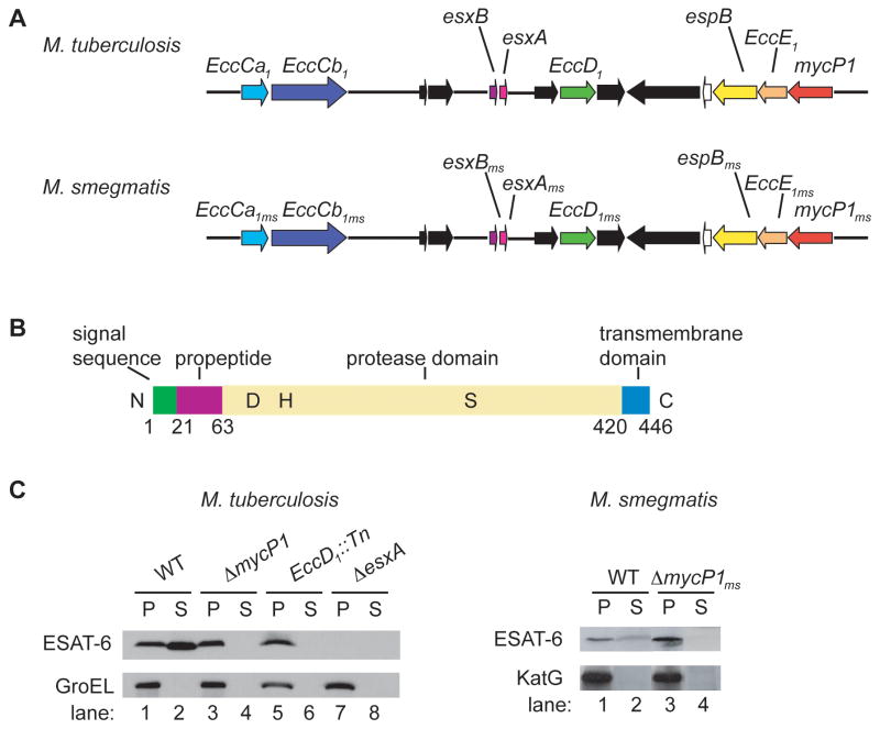
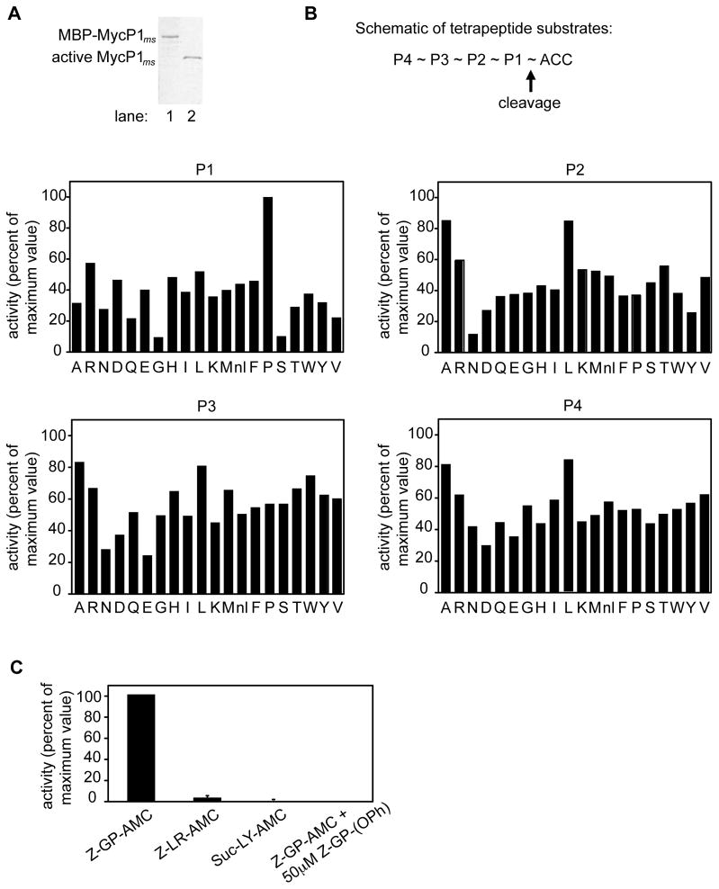
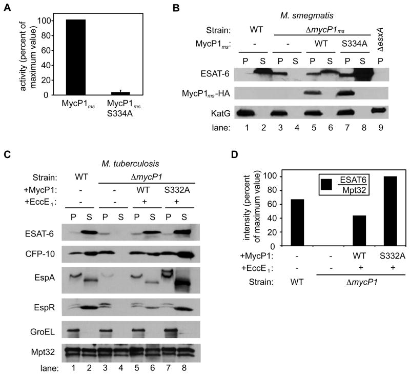
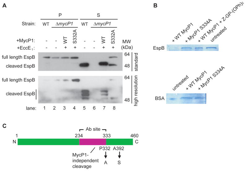
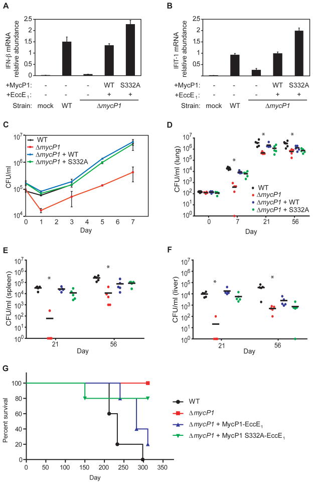
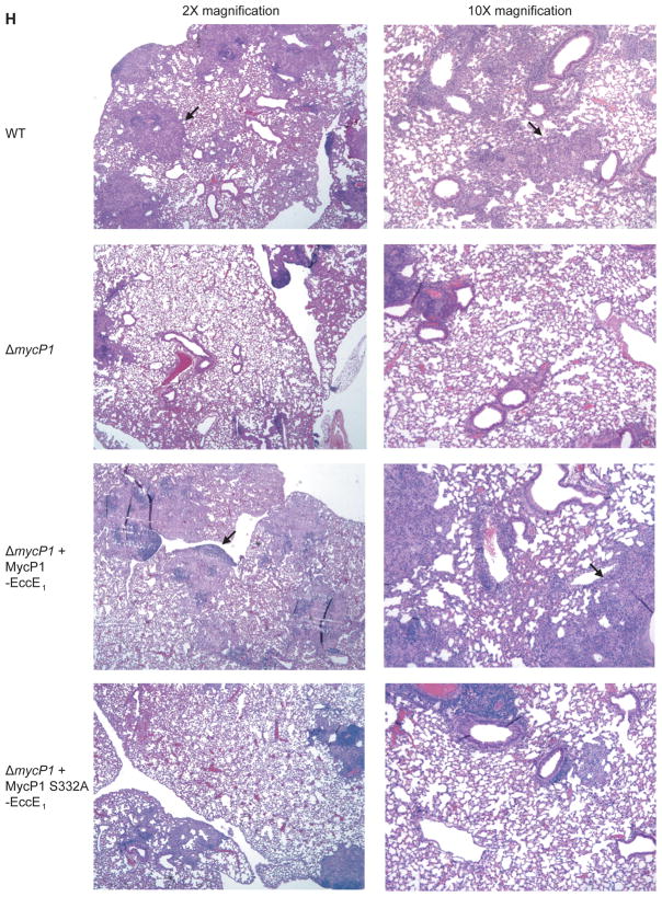
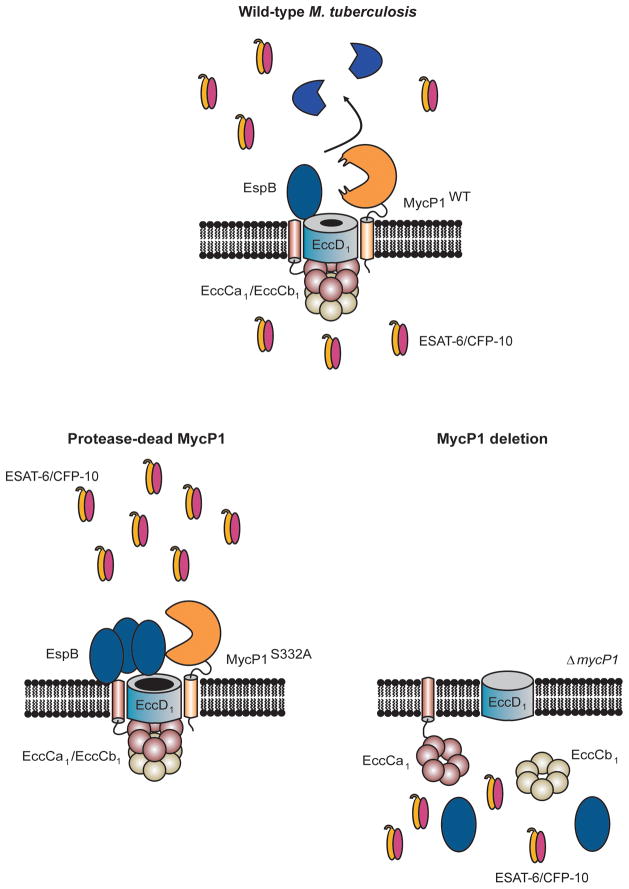
Mycobacterium tuberculosis uses the ESX-1 secretion system to deliver virulence proteins during infection of host cells. Here we report a mechanism of posttranscriptional control of ESX-1 mediated by MycP1, a M. tuberculosis serine protease. We show that MycP1 is required for ESX-1 secretion but that, unexpectedly, genetic inactivation of MycP1 protease activity increases secretion of ESX-1 substrates. We demonstrate that EspB, an ESX-1 substrate required for secretion, is a target of MycP1 in vitro and in vivo. During macrophage infection, an inactive MycP1 protease mutant causes hyperactivation of ESX-1-stimulated innate signaling pathways. MycP1 is required for growth in mice during acute infection, while loss of its protease activity leads to attenuated virulence during chronic infection. As the key ESX-1 substrates ESAT-6 and CFP-10 are highly immunogenic, fine-tuning of their secretion by MycP1 may balance virulence and immune detection and be essential for successful maintenance of long-term M. tuberculosis infection.


Figures

Figure 1
Figure 1. M. tuberculosis and M. smegmatis ΔmycP1 mutants fail to secrete ESAT-6
(A) Schematic representation of ESX-1 locus in M. tuberculosis and M. smegmatis. (B) Diagram of M. tuberculosis MycP1 showing domains and active site residues D90, H131 and S332. (C) Pellets (P) and cell culture supernatants (S) were generated from indicated strains and ESAT-6, GroEL and KatG were detected by Western blot. GroEL and KatG served as lysis controls. See Figure S1 for a description of the M. tuberculosis ΔmycP1 strain construction.
Figure 2
Figure 2. Purification and substrate specificity profiling of M. smegmatis MycP1
(A) MBP-MycP1 (lane 1) and active MycP1 (lane 2) were purified using amylose and hydroxyapatite affinity chromatography respectively, and resolved by SDS-PAGE. (B) MycP1 protease activity was measured against fluorogenic tetrapeptide substrate pools, in each of which the amino acid at one position was held constant while the other positions were varied. The x axis indicates which amino acid was held constant (nl = norleucine), while the y axis shows activity plotted relative to the highest activity of the library. (C) Activity of MycP1 against individual fluorogenic dipeptide substrates, in the absence or presence of specific inhibitor.
Figure 3
Figure 3. Protease activity of MycP1 negatively regulates ESX-1 secretion
(A) MycP1 protease activity was measured against the Z-GP-AMC substrate. Activity of protease-inactive MycP1 (MycP1 S334A), expressed relative to wild-type MycP1. (B) Western blot detection of ESAT-6, MycP1-HA and KatG in M. smegmatis pellets (P) and supernatants (S) from wild-type and mutant strains. (C) Western blot detection of ESAT-6, CFP-10, EspA, EspR, GroEL and Mpt32 in M. tuberculosis pellets (P) and supernatants (S) from wild-type and mutant cells. See Figure S2 for data showing that the ΔmycP1 mutation did not exert a polar effect on transcription of the downstream gene, eccE1. (D) Quantitation of ESAT6 in M. tuberculosis supernatants from wild-type and mutant strains, expressed relative to Sec-dependent substrate Mpt32.
Figure 4
Figure 4. MycP1 directly cleaves EspB, an ESX-1 substrate
(A) Western blot detection of EspB in M. tuberculosis pellets (P) and supernatants (S) from wild-type and mutant strains. The same samples were run on a standard 13 × 8 cm gel (W x L, top panel) and a higher resolution 16 × 16 cm gel (bottom panel) to reveal the different cleaved forms of EspB. (B) In vitro cleavage assay of MBP-EspB (top panel) and BSA (bottom panel) by WT MycP1, MycP1 S332A, and WT MycP1 in presence of specific inhibitor. (C) Schematic representation of EspB showing cleavage sites and the region used to generate the EspB antibody. See Figure S3 for MS results used to identify the EspB cleavage products.
Figure 5
Figure 5. Oversecretion of ESAT-6 by ΔmycP1 + MycP1 S332A mutant is sensed by macrophages, and MycP1 is required for virulence in macrophages and mice
Bone-marrow-derived macrophages were infected with wild-type and mutant strains of M. tuberculosis. At indicated time points total RNA was harvested from macrophage monolayers, and IFN-β (A) and IFIT-1 (B) mRNA levels were measured by quantitative PCR. Values were normalized to actin mRNA levels and each sample was assayed in triplicate. (C) Macrophages were infected, lysed at the indicated time points, and bacterial CFU were enumerated by plating. (D) BALB/c mice were infected with 102 CFU of each strain by aerosol, and bacteria were isolated from lungs immediately following infection, as well as at 7 and 21 days post infection. Five mice were used per time point and asterisks indicate significant differences by Kruskal-Wallis test. CFU were also obtained from spleens (E) and livers (F) of infected mice at 21 days post infection. (G) Survival of infected BALB/c mice (n = 5 per group). Statistical analysis indicated significant differences between all four strains (P<0.05) and more subtle differences between ΔmycP1 + WT MycP1 and ΔmycP1 + MycP1 S332A strains (P<0.13). (H) Hematoxylin/eosin-stained lung sections of mice infected with indicated strains, harvested 56 days post infection. Lungs from mice infected with wild-type and ΔmycP1 + WT MycP1 bacteria showed numerous, consolidated granulomas (arrows) while lungs from mice infected with ΔmycP1 and ΔmycP1 + MycP1 S332A strains showed fewer granulomas and more alveolar space. See Figures S4A and S4B for more images and results of our quantitative histopathology analysis.
Figure 6
Figure 6. Model of MycP1-mediated regulation of the ESX-1 secretion system in M. tuberculosis
In wild-type M. tuberculosis, MycP1 is part of a complex and proteolyzes EspB in the periplasm. Full-length EspB is an activator of ESX-1 secretion and its cleavage functions to inhibit secretion. In ΔmycP1 + MycP1 S332A cells, full-length EspB accumulates and therefore ESX-1 secretion is hyper-activated. In ΔmycP1 bacteria, secretion is abolished because the secretion complex fails to assemble in the absence of MycP1.

Comment in

  • The two faces of MycP1.
    David R. David R. Nat Rev Microbiol. 2010 May;8(5):314. doi: 10.1038/nrmicro2360. Nat Rev Microbiol. 2010. PMID: 21080601 No abstract available.

References

    1. Abdallah AM, Gey van Pittius NC, Champion PAD, Cox JS, Luirink J, Vandenbroucke-Grauls CMJE, Appelmelk BJ, Bitter W. Type VII secretion – mycobacteria show the way. Nature Reviews. 2007;5:883–891. - PubMed
    1. Ando M, Yoshimatsu T, Ko C, Converse PJ, Bishai WR. Deletion of Mycobacterium tuberculosis sigma factor E results in delayed time to death with bacterial persistence in the lungs of aerosol-infected mice. Infect Immun. 2003;71:7170–7172. - PMC - PubMed
    1. Bergeron F, Leduc R, Day R. Subtilase-like pro-protein convertases: from molecular specificity to therapeutic applications. J Mol Endocrinol. 2000;24:1–22. - PubMed
    1. Bitter W, Houben EN, Bottai D, Brodin P, Brown EJ, Cox JS, Derbyshire K, Fortune SM, Gao LY, Liu J, Gey van Pittius NC, Pym AS, Rubin EJ, Sherman DR, Cole ST, Brosch R. Systematic genetic nomenclature for type VII secretion systems. PLoS Pathog. 2009;5:e1000507. - PMC - PubMed
    1. Brandt L, Elhay M, Rosenkrands I, Lindblad EB, Andersen P. ESAT-6 subunit vaccination against Mycobacterium tuberculosis. Infect Immun. 2000;68:791–795. - PMC - PubMed

Publication types

MeSH terms

LinkOut - more resources