. 2010 Mar 18;7(3):221-33.
doi: 10.1016/j.chom.2010.02.007.

Flexible use of nuclear import pathways by HIV-1

Affiliations

Flexible use of nuclear import pathways by HIV-1

KyeongEun Lee et al. Cell Host Microbe. .

Abstract

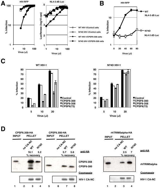
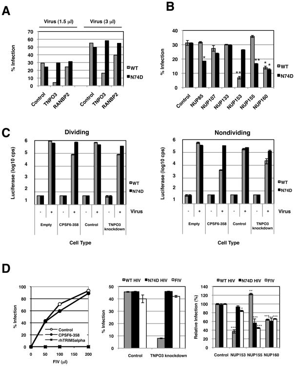
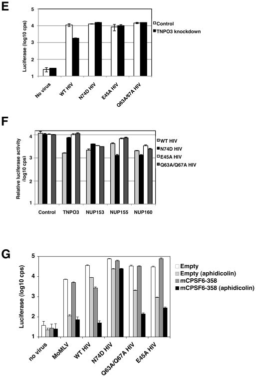
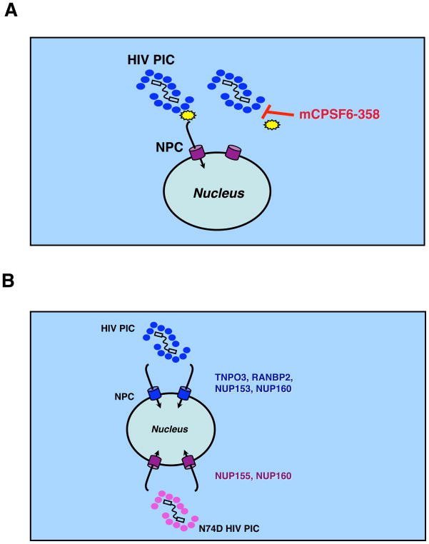
HIV-1 replication requires transport of nascent viral DNA and associated virion proteins, the retroviral preintegration complex (PIC), into the nucleus. Too large for passive diffusion through nuclear pore complexes (NPCs), PICs use cellular nuclear transport mechanisms and nucleoporins (NUPs), the NPC components that permit selective nuclear-cytoplasmic exchange, but the details remain unclear. Here we identify a fragment of the cleavage and polyadenylation factor 6, CPSF6, as a potent inhibitor of HIV-1 infection. When enriched in the cytoplasm, CPSF6 prevents HIV-1 nuclear entry by targeting the viral capsid (CA). HIV-1 harboring the N74D mutation in CA fails to interact with CPSF6 and evades the nuclear import restriction. Interestingly, whereas wild-type HIV-1 requires NUP153, N74D HIV-1 mimics feline immunodeficiency virus nuclear import requirements and is more sensitive to NUP155 depletion. These findings reveal a remarkable flexibility in HIV-1 nuclear transport and highlight a single residue in CA as essential in regulating interactions with NUPs.


Figures

Figure 1
Figure 1
Alignment of the predicted open reading frames of wild-type human CPSF6, wild-type mouse CPSF6 (68 kD splice variant), and mCPSF6-358. Human residues encoded by exon 6 are shown, though, as indicated for mCPSF6, this stretch is missing from the 68 kD form. Exon boundaries and protein domains are denoted by downward triangles and shaded boxes, respectively. Protein domains including the RNA-recognition motif (RRM) are labeled (Ruegsegger et al., 1998), and NLS determinants are boxed (Dettwiler et al., 2004). Amino acids in italics are acquired from the pMIGR1 vector and not needed for antiviral function (not shown).
Figure 2
Figure 2. C-terminally truncated CPSF6 restricts HIV-1 infection
(A) Infection of NIH3T3.hCycT1 cells stably expressing different CPSF6 molecules by HIV-RFP/VSV-G. (B) Upper panels: Subcellular localization of endogenous, ectopically-expressed wild-type, and mutant CPSF6 molecules in NIH3T3.hCycT1 cell lines that express the mutant proteins. Western blot analysis was done with polyclonal antisera to determine the distribution of CPSF6 proteins in cells that contain only the empty MIGR1 vector versus those that express mCPSF6-358, wild-type mCPSF6(68), or the minimally truncated mCPSF6(68)-526. Lower panels: The blots were reprobed with tubulin and histone antibodies to validate cell fractionation. (C) mCPSF6-358 blocks HIV-1 infection in primary T cells. CD4+ T cells activated through the TCR by using anti-CD3 and anti-CD28 as previously described (Unutmaz et al., 1999) were transduced with lentiviral vectors expressing IRES-HSA, rhTRIM5alpha-IRES-HSA, or mCPSF6-358-IRES-HSA. The infected cells were expanded in IL-2 containing media for 4 days and superinfected at different multiplicities of infection (m.o.i.) by HIV-eGFP/VSV-G. Numbers represent % of GFP+ cells over HSA+ cells day 4 post superinfection. (D) Infection of HeLa cells expressing mCPSF6-358 with HIV-RFP/VSV-G and MX-RFP/VSV-G vectors. The percent of the cells that were infected was measured by FACS for RFP expression; the amounts of the virus containing supernatants is shown on the x-axis. (E) Replication competent primate lentiviruses are inhibited by mCPSF6-358. Infections were performed under single-cycle conditions using MAGI cells expressing CCR5. Cells that expressed mCPSF6-358 are shown by the black bars. MLV expressing HIV-1 Tat and pseudotyped with VSV-G was used as a control.
Figure 3
Figure 3. Analysis of HIV-1 replication in the presence of truncated CPSF6
(A) Total HIV-1 DNA is reduced in NIH3T3.hCycT1 cells expressing mCPSF6-358. Left: 3 d post-infection with HIV-luc/VSV-G, a portion of infected cells were analyzed for luciferase activity. Right: 7 d post-infection, DNA extracted from cells was analyzed by qPCR using gag-specific primers. Viral DNA copies per microgram genomic DNA are depicted on the y-axis. (B) qPCR measurements of HIV-1 DNA synthesis. 293T cells expressing MIGR1-mCPSF6 or MIGR1-mCPSF6-358 and infected with HIV-HSA/VSV-G or HIVIN/D116N-HSA/VSV-G carrying an active site mutation in the viral IN (Engelman et al., 1995) were lysed at various times to measure the amount of viral DNA using primers for specific steps in reverse transcription. Early (minus strand), late (plus strand), and 2-LTR circle vDNA forms are labeled on the tops of the graphs. (C) Late reverse transcripts and PIC activity from NIH3T3.hCycT1 cells expressing mCPSF6-358. NIH3T3/MIGR1 or NIH3T3/MIGR1-mCPSF6-358 cells infected with HIV-luc/VSV-G were lysed at 9 h post-infection. Late reverse transcripts were measured by qPCR as shown in the left graph, and the amount of PIC activity normalized to the levels of late reverse transcription products in the different samples is shown on the right. The activity of PICs extracted from empty vector control cells was arbitrarily set at 100%. Error bars indicate the standard errors of means derived from duplicate integration assays.
Figure 4
Figure 4. A mutation in HIV-1 CA confers resistance to mCPSF6-358
(A) Selection for HIV-1 resistance to mCPSF6-358. HUT-R5 cells that contain the empty vector or HUT-R5.mCPSF6-358 cells were infected with HIV-1NL4-3/BaL and passaged every 2–3 days. Culture supernatants were collected and analyzed for CA by HIV-1 p24 ELISA. (B) HIV-1 with the N74D mutation in CA efficiently infects nondividing HeLa cells and infection is not blocked in nondividing HeLa cells expressing mCPSF6-358. HeLa.MIGR1 and HeLa.MIGR1-mCPSF6-358 cells were maintained in the presence or absence of aphidicolin and infected with MLV, HIVNLdE-luc/VSV-G, or HIVNLdE CA N74D-luc/VSV-G. Two days after infection, cells were lysed and assayed for luciferase activity.
Figure 5
Figure 5. CPSF6-358 interacts with wild-type HIV-1 CA
(A) Different HIV-1 vectors can saturate the CPSF6-358 restriction. 293T.LPCX (control) or 293T.LPC-CPSF6-358-HA cell populations were challenged with increasing volumes of WT vs. N74D HIV-RFP/VSV-G or luciferase vectors (HIVNLdE-luc/VSV-G or HIVNLdE CA N74D-luc/VSV-G). Infectivity was measured by FACS or luciferase activity. (B) Pre-infection with WT HIV-1 abrogates the CPSF6-358 restriction to a second virus. 293T.LPC-CPSF6-358-HA cells were challenged with increasing volumes of HIVNLdE-luc/VSV-G or HIVNLdE CA N74D-luc/VSV-G and then with a fixed amount of WT HIV-RFP/VSV-G. Infectivity by WT HIV-RFP/VSV-G was measured by FACS. (C) CPSF6-300 does not restrict WT HIV-1. WT vs. N74D HIV-RFP/VSV-G infection of 293T cells transiently transfected with empty vector (control) or HA-tagged constructs expressing human CPSF6-300, CPSF6-358, or CPSF6 (encoding the full length 72 kD isoform). The percent of the cells that were infected was measured by FACS for RFP expression; the amounts of the virus containing supernatants is shown on the x-axis. (D) Enhanced binding of CPSF6-358 to WT CA-NC complexes. Recombinant WT and N74D CA-NC complexes were incubated with lysates from 293T cells transfected with HA-tagged constructs expressing CPSF6-300, CPSF6-358, or rhTRIM5alpha. Mixtures were then layered on top of a 70% sucrose cushion followed by ultracentrifugation. Top panels are Western blots using anti-HA; bottom panels are Coomassie stains of pelleted material. Lanes 1 and 5 are lysate input lanes that were not subjected to ultracentrifugation. Lanes 2 and 6 are pelleted material in the absence of CA-NC complexes. Recovery of HA-tagged proteins relative to input was measured by densitometry.
Figure 6
Figure 6. WT and N74D HIV-1 require different nuclear pore factors
(A) N74D HIV-1 vectors do not require TNPO3 or RANBP2 for infection. HeLa cells were transiently transfected with control siRNA oligos and with oligos directed at TNPO3 or RANBP2. One day after siRNA transfection, cells were replated for virus challenge. Two days after siRNA transfection, the transfected cells were infected with 1.5 and 3.0 μl of WT or N74D HIV-RFP/VSV-G and assayed 48 h later by FACS. (B) Differential NUP requirements by WT HIV-1 and the N74D mutant. HeLa cells were transfected with siRNAs that target different NUP genes (NUP85, NUP107, NUP133, NUP153, NUP155, and NUP160). Nontargeting siRNA was also transfected as a control. Infections with WT or N74D HIV-RFP/VSV-G were performed in duplicate and assayed 48 h later by FACS. Statistical analysis was performed by Student’s t test; *p < 0.05 and **p <0.01 versus WT or N74D HIV-1 infection of respective control infected cells. (C) Cell growth arrest does not increase the dependence of WT HIV-1 on TNPO3. HeLa cells that contain only the empty vector or cells that express mCPSF6-358 and HeLa cells transduced with control shRNA vector or TNPO3 shRNA were infected with either WT HIV-1 or N74D mutant vectors that express a luciferase reporter (HIVNLdE-luc/VSV-G or HIVNLdE CA N74D-luc/VSV-G, respectively). Cell lysates were measured for luciferase activity 48 h after infection. Luciferase values reflect averages of duplicate infections with standard deviations. The data in the right panel were from cells treated with aphidicolin. (D) N74D HIV-1 is an FIV phenocopy. Left panel, 293T cells stably expressing BabePuro (empty vector control), BabePuro-mCPSF6-358, or LPC-rhTRIMalpha were infected with FIV-GFP(GinSin)/VSV-G (Loewen et al., 2003). The percentage of infected cells was measured by FACS for GFP expression; the amounts of the virus containing supernatants is shown on the x-axis. Middle panel, HeLa.control_shRNA or HeLa.TNPO3_shRNA cells were infected in duplicates with WT HIV-RFP/VSV-G, N74D HIV-RFP/VSV-G, or FIV-GFP/VSV-G. Infection was measured by FACS. Right panel, HeLa cells transiently transfected with nontargeting and NUP targeting siRNA were infected in duplicates with WT HIV-RFP/VSV-G, N74D HIV-RFP/VSV-G, or FIV-GFP/VSV-G. The average of four independent experimental trials is depicted as a relative infection value. Statistical analysis was performed by Student’s t test; *p = 0.0205, **p = 0.0009, and ***p < 0.0001 relative WT or N74D HIV-1 infection of respective control infected cells. (E) E45A and Q63A/Q67A HIV-1 are TNPO3-independent. HeLa cells stably transduced with control shRNA or TNPO3 shRNA vectors were infected with WT HIV-1 or different CA mutant HIV-1 (HIVNLdE-luc/VSV-G, HIVNLdE CA N74D-luc/VSV-G, HIVNLdE CA E45A-luc/VSV-G, or HIVNLdE CA Q63A/Q67A-luc/VSV-G). After 48 h of infection, cell lysates were collected to measure luciferase activity. Luciferase values reflect averages of duplicate infections with standard deviations. (F) E45A and Q63A/Q67A HIV-1 are less sensitive to NUP depletions. HeLa cells were transiently transfected with nontargeting siRNA or siRNAs that target different host factors (TNPO3, NUP153, NUP155, and NUP160). Cells were infected with HIVNLdE-luc/VSV-G, HIVNLdE CA N74D-luc/VSV-G, HIVNLdE CA E45A-luc/VSV-G, or HIVNLdE CA Q63A/Q67A-luc/VSV-G and lucifease activity from lysates was measured 48 h later. Control cell virus infections are within 10-fold of their actual activities and were used to normalize infection values of other samples. (G) CPSF6-358 restriction of E45A or Q63A/Q67A HIV-1 is cell-cycle dependent. HeLa.MIGR1 and HeLa.MIGR1-mCPSF6-358 cells maintained in the presence or absence of aphidicolin were infected in duplicate with MLV, HIVNLdE-luc/VSV-G, HIVNLdE CA N74D-luc/VSV-G, HIVNLdE CA E45A-luc/VSV-G, or HIVNLdE CA Q63A/67A-luc/VSV-G. Cells were lysed and assayed for luciferase activity two days after infection. Error bars represent standard deviations of duplicates.
Figure 7
Figure 7. Models for HIV-1 nuclear entry
(A) HIV-1 nuclear entry is restricted by CPSF6-358 interaction with the CA core (blue ovals), thus interfering with HIV-1 core disassembly or interaction with a host transport factor (yellow polygon). (B) HIV-1 CA regulates nuclear transport and pore protein requirements for infection. Depletion of TNPO3, RANBP2, and NUP153 diminishes WT but not N74D HIV-1 infection. By contrast, depletion of NUP155 limits N74D HIV-1 and FIV infection. Knockdown of NUP160 impaired infection by different lentiviruses.

References

    1. Arhel NJ, Souquere-Besse S, Munier S, Souque P, Guadagnini S, Rutherford S, Prevost MC, Allen TD, Charneau P. HIV-1 DNA Flap formation promotes uncoating of the pre-integration complex at the nuclear pore. EMBO J. 2007;26:3025–3037. - PMC - PubMed
    1. Boehmer T, Jeudy S, Berke IC, Schwartz TU. Structural and functional studies of Nup107/Nup133 interaction and its implications for the architecture of the nuclear pore complex. Mol Cell. 2008;30:721–731. - PMC - PubMed
    1. Bouyac-Bertoia M, Dvorin JD, Fouchier RA, Jenkins Y, Meyer BE, Wu LI, Emerman M, Malim MH. HIV-1 infection requires a functional integrase NLS. Mol Cell. 2001;7:1025–1035. - PubMed
    1. Brass AL, Dykxhoorn DM, Benita Y, Yan N, Engelman A, Xavier RJ, Lieberman J, Elledge SJ. Identification of host proteins required for HIV infection through a functional genomic screen. Science. 2008;319:921–926. - PubMed
    1. Bukrinsky M. A hard way to the nucleus. Mol Med. 2004;10:1–5. - PMC - PubMed

Publication types