. 2010 Mar 18;7(3):234-44.
doi: 10.1016/j.chom.2010.02.005.

The Cellular lysine methyltransferase Set7/9-KMT7 binds HIV-1 TAR RNA, monomethylates the viral transactivator Tat, and enhances HIV transcription

Affiliations

The Cellular lysine methyltransferase Set7/9-KMT7 binds HIV-1 TAR RNA, monomethylates the viral transactivator Tat, and enhances HIV transcription

Sara Pagans et al. Cell Host Microbe. .

Abstract

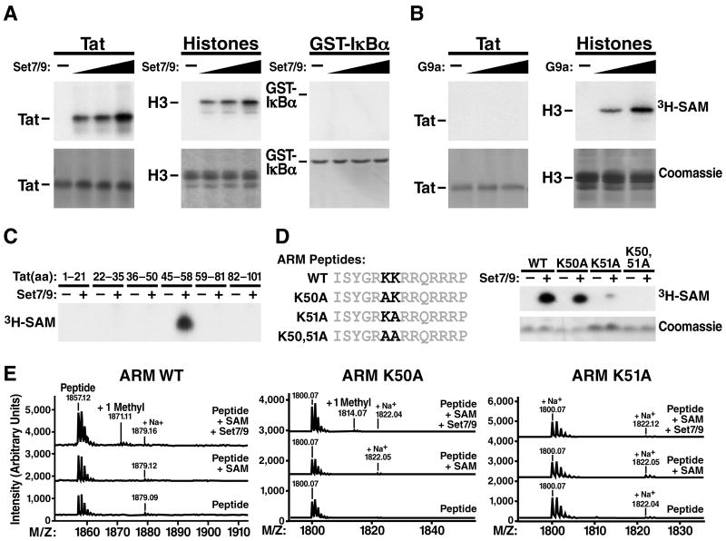
The Tat protein of HIV-1 plays an essential role in HIV gene expression by promoting efficient elongation of viral transcripts. Posttranslational modifications of Tat fine-tune interactions of Tat with cellular cofactors and TAR RNA, a stem-loop structure at the 5' ends of viral transcripts. Here, we identify the lysine methyltransferase Set7/9 (KMT7) as a coactivator of HIV transcription. Set7/9-KMT7 associates with the HIV promoter in vivo and monomethylates lysine 51, a highly conserved residue located in the RNA-binding domain of Tat. Knockdown of Set7/9-KMT7 suppresses Tat transactivation of the HIV promoter, but does not affect the transcriptional activity of methylation-deficient Tat (K51A). Set7/9-KMT7 binds TAR RNA by itself and in complex with Tat and the positive transcription elongation factor P-TEFb. Our findings uncover a positive role for Set7/9-KMT7 and Tat methylation during early steps of the Tat transactivation cycle.


Figures

Figure 1
Figure 1. In Vitro Methylation of Tat by Set7/9-KMT7
(A) Synthetic Tat (72 aa), histones or recombinant GST-IκBα proteins were incubated with 3H-radiolabeled S-adenosyl-methionine (SAM) and increasing amounts of recombinant Set7/9-KMT7 (0, 0.5, 1, or 2 μg). Tat and histone H3 methylation were visualized by autoradiography (top panels). (B) Radioactive in vitro methylation reactions with synthetic Tat or histones and recombinant G9a enzyme (0, 1, or 2 μg). (C) In vitro methylation assays performed with short Tat peptides, recombinant Set7/9-KMT7, and 3H-SAM. Peptides were separated on Tris-Tricine gels and visualized by autoradiography. (D) In vitro methylation assays of ARM peptides (aa 45–58), containing either two lysines (WT) or alanine substitutions at positions K50 and K51. (E) MALDI TOF mass spectrometry of nonradioactive methylation reactions performed with the wild-type (WT), K50A or K51A ARM peptides. Peptides were incubated with Set7/9-KMT7 and SAM, SAM alone or only the reaction buffer. (See also Figure S1).
Figure 2
Figure 2. Tat Is Monomethylated at K51 in Cells
(A) Dot blot analysis of biotinylated ARM peptides, either unmodified, mono-, di-, or trimethylated at K51 with α-meARM antibodies or SA-HRP. (B) Western blot analysis of in vitro methylation reactions with biotinylated synthetic Tat (12.5, 25, 50 and 100 ng), recombinant Set7/9-KMT7 and nonradioactive SAM with α-meARM antibodies or SA-HRP. (C) Immunoprecipitations of wild-type or mutant K51A Tat/FLAG in 293 cells, followed by western blotting with α-meARM or α-FLAG antibodies. (D) Immunoprecipitation/western blot analysis of Tat/FLAG coexpressed with Set7/9-KMT7 in 293 cells. The α-meARM antibodies were preincubated with milk, a 10× molar excess of K51-monomethylated ARM peptide or a 10× molar excess of non-modified ARM peptide. (E) Immunoprecipitation of Tat/FLAG in Jurkat A2 cells treated with TNF-α followed by western blotting with α-meARM or α-FLAG antibodies.
Figure 3
Figure 3. Set7/9-KMT7 Activates Tat Transactivation through K51 Methylation
(A) Cotransfections in HeLa cells of expression vectors for wild-type or catalytically inactive Set7/9-KMT7 (H297A; 150 ng), with the indicated amounts of Tat expression vector and the HIV LTR luciferase reporter (200 ng). In parallel, cotransfections were performed with the EF-1α-RL reporter (20 ng) and wild-type or catalytically inactive Set7/9-KMT7 (H297A; 150 ng). Luciferase or Renilla values were analyzed 24 h after transfections. The average of three independent experiments (mean ± SEM) is shown; * corresponds to a p value <0.05 and ** to a p value <0.01 compared to cells transfected with vector control. (B) SiRNA-mediated knockdown of Set7/9-KMT7 in HeLa cells. Cells were cotransfected with the HIV LTR luciferase construct (200 ng) and increasing amounts of expression vectors for wild-type or mutant K51A Tat/FLAG (0, 10, 50, 250 ng) 48 h after siRNA transfection. Measurements of luciferase activity and western blotting were performed 24 h after plasmid transfections. Luciferase values represent the average (mean ± SEM) of three experiments; * corresponds to a p value <0.05 and ** to a p value <0.01 compared to control cells expressing wild-type Tat. (C) Transcriptional activity of TatK51A and TatK51R mutants in Set7/9-KMT7 knockdown cells. Experiment was performed as in (B) with 250 ng of wild-type or mutant FLAG-Tat. Luciferase values represent the average (mean ± SEM) of three experiments. * corresponds to a p value <0.05 compared to wild-type Tat-transfected cells.
Figure 4
Figure 4. Set7/9-KMT7 Regulates HIV Gene Expression in the Context of Lentiviral Infection
(A) SiRNA-mediated knockdown of Set7/9-KMT7 in latently infected Jurkat A2 cells. Three days after nucleofection of Set7/9-KMT7 or control siRNAs, western blotting was performed, and cells were stimulated with PMA for 12 h or were left unstimulated. GFP expression was measured by flow cytometry. The average (mean ± SEM) of three independent experiments is shown, * corresponds to a p value <0.05 compared to control siRNA-transfected cells. (B) ShRNA-mediated knockdown of Set7/9-KMT7 in CD4+T cells. Primary CD4+ T cells isolated from uninfected blood donors were infected first with lentiviral particles encoding shRNAs against Set7/9-KMT7, and second with a GFP-tagged reporter virus in the background of HIVNL4-3 or a lentiviral vector expressing GFP from the EF-1α promoter. GFP expression within shRNA-expressing cells (as marked by mCherry expression) is expressed relative to cells expressing non-targeting shRNA control. Average (mean ± SD) from triplicate experiments performed with three different donors (HIV GFP) or a single donor (HIV (EF-1α) GFP) is shown. Western blotting was performed in Jurkat cells transduced with indicated shRNAs and sorted for mCherry expression. (See also Figure S2).
Figure 5
Figure 5. In Vivo Recruitment of Set7/9-KMT7 to the HIV LTR
(A) Chromatin immunoprecipitation analysis of Set7/9-KMT7 in Jurkat A2 cells treated with PMA for 4 h or left unstimulated. Real-time PCR was used to quantify the enrichment of indicated DNA regions after immunoprecipitation with α-Set7/9-KMT7 antibodies. Quantities of immunoprecipitated DNA were normalized to input DNA and expressed relative to the β-actin control. Three independent experiments were performed with similar outcome. Average (mean ± SD) of three PCR reactions of one experiment is shown. (B) Coimmunoprecipitation of Tat/FLAG and endogenous Set7/9-KMT7 in Jurkat A2 cells treated with TNF-α. Cellular lysates were immunoprecipitated with α-Set7/9-KMT7 antibodies followed by western blotting with α-Set7/9-KMT7 or α-FLAG antibodies. (C) Coimmunoprecipitation of wild-type and K51A-mutant Tat with endogenous Set7/9-KMT7 in 293 cells. Immunoprecipitations were performed with α-Set7/9-KMT7 antibodies or agarose beads alone and analyzed by western blotting with α-Set7/9-KMT7 or α-FLAG antibodies. (See also Figure S3).
Figure 6
Figure 6. Set7/9-KMT7 Binds TAR RNA and Tat/P-TEFb
(A) Gel shift assays of recombinant Set7/9-KMT7 (0, 50, 150 and 500 ng) and radiolabeled wild-type (WT) or bulge-mutant (ΔBulge) TAR RNA probes. A 10× excess of non-radiolabeled TAR RNA was included to compete for binding with the radiolabeled probe. (See also Figure S4A). (B) Supershift assays with α-Set7/9-KMT7 antibodies (7 μg) or control rabbit IgGs in reactions containing Set7/9-KMT7 (1 μg) and radiolabeled wild-type TAR RNA probe. (C) Gel shift assays with full-length or N-terminally truncated Set7/9-KMT7 (0, 0.1, 0.3, 1 μg) and radiolabeled wild-type TAR RNA probe. (D) Tat and P-TEFb binding to TAR in the presence of Set7/9-KMT7. Radiolabeled TAR RNA probes (WT or ΔLoop) were incubated with synthetic Tat protein (0, 2.5, 10 and 40 ng) in the absence or presence of recombinant Set7/9-KMT7 (500 ng). The same reactions were performed in the presence of recombinant Cyclin T1/CDK9 (400 ng). (E) Coimmunoprecipitation of Cyclin T1 and Set7/9-KMT7 in the presence of Tat. 293 cells were cotransfected with HA-Cyclin T1 and Tat/FLAG expression vectors. Cell lysates were immunoprecipitated with α-Set7/9-KMT7 antibodies followed by western blotting with α-Set7/9-KMT7, α-FLAG and α-Cyclin T1 antibodies. (See also Figure S4B).
Figure 7
Figure 7. Model of Set7/9-KMT7 Recruitment to the HIV LTR
(A) Set7/9-KMT7 binds TAR bulge and loop sequences in newly synthesized HIV transcripts. (B) When Tat is produced, Set7/9-KMT7 engages into complex formation with Tat and P-TEFb and methylates K51 in Tat. See text for details.

Comment in

  • The HIV-1 Tat team gets bigger.
    Rice AP. Rice AP. Cell Host Microbe. 2010 Mar 18;7(3):179-81. doi: 10.1016/j.chom.2010.03.001. Cell Host Microbe. 2010. PMID: 20227660

Similar articles

Cited by

References

    1. Agbottah E, Deng L, Dannenberg LO, Pumfery A, Kashanchi F. Effect of SWI/SNF chromatin remodeling complex on HIV-1 Tat activated transcription. Retrovirology. 2006;3:48. - PMC - PubMed
    1. Anand K, Schulte A, Vogel-Bachmayr K, Scheffzek K, Geyer M. Structural insights into the cyclin T1-Tat-TAR RNA transcription activation complex from EIAV. Nat Struct Mol Biol. 2008;15:1287–1292. - PubMed
    1. Ariumi Y, Serhan F, Turelli P, Telenti A, Trono D. The integrase interactor 1 (INI1) proteins facilitate Tat-mediated human immunodeficiency virus type 1 transcription. Retrovirology. 2006;3:47. - PMC - PubMed
    1. Barboric M, Peterlin BM. A new paradigm in eukaryotic biology: HIV Tat and the control of transcriptional elongation. PLoS Biol. 2005;3:e76. - PMC - PubMed
    1. Benkirane M, Chun RF, Xiao H, Ogryzko VV, Howard BH, Nakatani Y, Jeang KT. Activation of integrated provirus requires histone acetyltransferase. p300 and P/CAF are coactivators for HIV-1 Tat. J Biol Chem. 1998;273:24898–24905. - PubMed

Publication types

MeSH terms

Substances

LinkOut - more resources