

Urothelium-derived Sonic hedgehog promotes mesenchymal proliferation and induces bladder smooth muscle differentiation
- PMID: 20227816
- PMCID: PMC3712847
- DOI: 10.1016/j.diff.2010.02.002
Urothelium-derived Sonic hedgehog promotes mesenchymal proliferation and induces bladder smooth muscle differentiation
Abstract
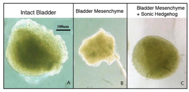
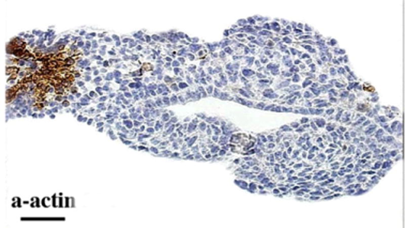
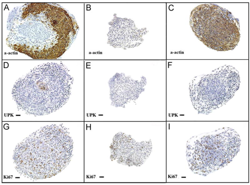
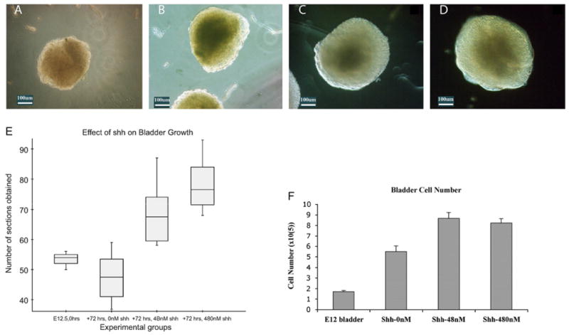
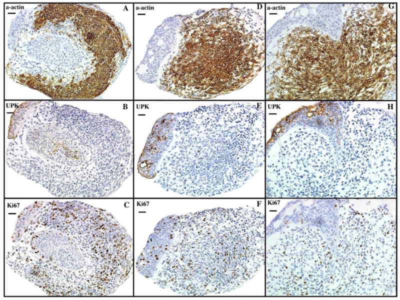
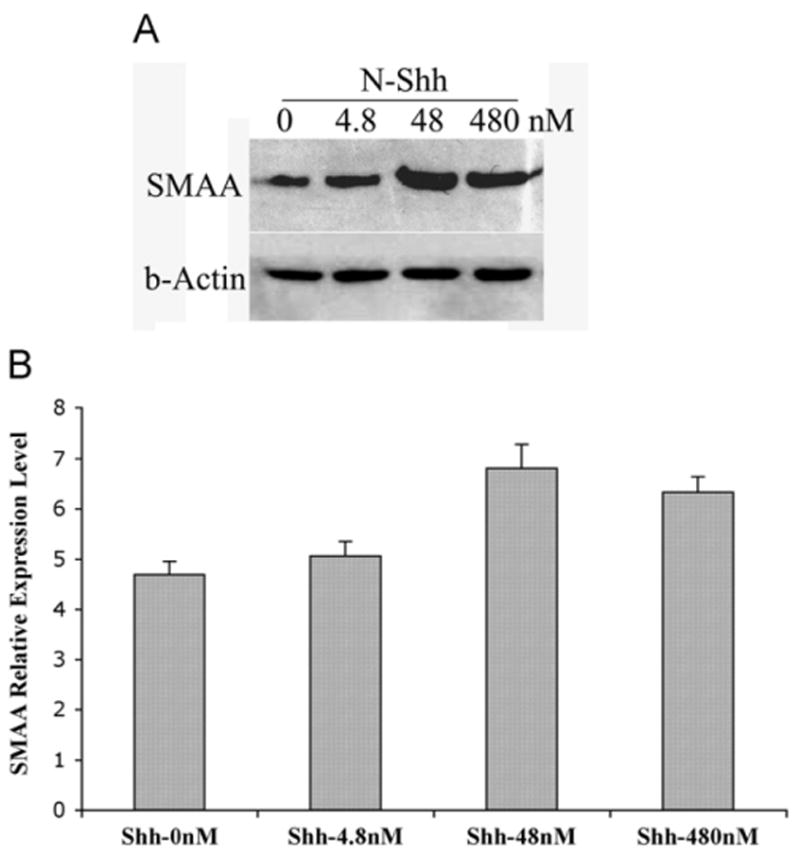
Induction of smooth muscle differentiation from bladder mesenchyme depends on signals that originate from the urothelium. We hypothesize Sonic hedgehog (Shh) is the urothelial signal that promotes bladder mesenchymal proliferation and induces bladder smooth muscle differentiation. Pregnant FVB mice were euthanized on embryonic day (E) 12.5 and fetal bladders were harvested. Two experimental protocols were utilized: Specimens were sized by serial sectioning. Cell counts were performed after trypsin digestion. Immunohistochemistry was performed to detect smooth muscle-specific protein expression. alpha-Actin expression was quantified using Western blot. All specimens were viable at 72h. BLM cultured without Shh survived but did not grow or undergo smooth muscle differentiation. IB cultured without Shh and BLM cultured with Shh grew and expressed smooth muscle proteins at 72h. IB cultured with Shh were larger and contained more cells than IB cultured without Shh (all p<0.05). Increasing Shh concentration from 48 to 480nM did not change bladder size, cell counts, or the level of alpha-actin expression. Prior to culture, IB did not express alpha-actin. After culture of IB in Shh-deficient media, alpha-actin was detected throughout the mesenchyme except in the submucosal layer. The IB submucosa was thinner after culture with 48nM Shh and smooth muscle completely obliterated the submucosa after culture with 480nM Shh. In fetal mouse bladders, urothelium-derived Shh is necessary for mesenchymal proliferation and smooth muscle differentiation. Shh concentration affects mesenchymal proliferation and patterning of bladder smooth muscle.
Figures

Similar articles
-
Smooth muscle differentiation and patterning in the urinary bladder.Differentiation. 2010 Sep-Oct;80(2-3):106-17. doi: 10.1016/j.diff.2010.05.004. Epub 2010 Jun 12. Differentiation. 2010. PMID: 20541860 Free PMC article. Review.
-
Urothelial sonic hedgehog signaling plays an important role in bladder smooth muscle formation.Differentiation. 2007 Dec;75(10):968-77. doi: 10.1111/j.1432-0436.2007.00187.x. Epub 2007 May 9. Differentiation. 2007. PMID: 17490411
-
Role of mesenchymal-epithelial interactions in normal bladder development.J Urol. 1996 Nov;156(5):1820-7. J Urol. 1996. PMID: 8863624
-
Signalling molecules involved in mouse bladder smooth muscle cellular differentiation.Int J Dev Biol. 2010;54(1):175-80. doi: 10.1387/ijdb.082610bl. Int J Dev Biol. 2010. PMID: 20013655 Free PMC article.
-
Cellular signaling in the bladder.Front Biosci. 1997 Dec 1;2:d592-5. doi: 10.2741/a215. Front Biosci. 1997. PMID: 9374449 Review.
Cited by
-
Smooth muscle differentiation and patterning in the urinary bladder.Differentiation. 2010 Sep-Oct;80(2-3):106-17. doi: 10.1016/j.diff.2010.05.004. Epub 2010 Jun 12. Differentiation. 2010. PMID: 20541860 Free PMC article. Review.
-
Epithelial and mesenchymal compartments of the developing bladder and urethra display spatially distinct gene expression patterns.Dev Biol. 2025 Apr;520:155-170. doi: 10.1016/j.ydbio.2025.01.005. Epub 2025 Jan 9. Dev Biol. 2025. PMID: 39798644 Free PMC article.
-
Fgfr2 is integral for bladder mesenchyme patterning and function.Am J Physiol Renal Physiol. 2015 Apr 15;308(8):F888-98. doi: 10.1152/ajprenal.00624.2014. Epub 2015 Feb 4. Am J Physiol Renal Physiol. 2015. Retraction in: Am J Physiol Renal Physiol. 2016 Jul 1;311(1):F239. doi: 10.1152/ajprenal.zh2-7964-retr.2016. PMID: 25656370 Free PMC article. Retracted.
-
Interplay of SHH, WNT and BMP4 signaling regulates the development of the lamina propria in the murine ureter.Development. 2025 Feb 1;152(3):DEV204214. doi: 10.1242/dev.204214. Epub 2025 Feb 6. Development. 2025. PMID: 39817691 Free PMC article.
-
Mechanisms and implications of epithelial cell plasticity in the bladder.Nat Rev Urol. 2025 Jul 24. doi: 10.1038/s41585-025-01066-y. Online ahead of print. Nat Rev Urol. 2025. PMID: 40707815 Review.
References
-
- Baskin LS, Hayward SW, Sutherland RA, DiSandro MJ, Thomson AA, Goodman J, Cunha GR. Mesenchymal–epithelial interactions in the bladder. World J Urol. 1996;14:301–309. - PubMed
-
- Bitgood MJ, McMahon AP. Hedgehog and Bmp genes are coexpressed at many diverse sites of cell–cell interaction in the mouse embryo. Dev Biol. 1995;172:126–138. - PubMed
Publication types
MeSH terms
Substances
Grants and funding
LinkOut - more resources
Full Text Sources
Molecular Biology Databases
