. 2010 May 28;285(22):16757-70.
doi: 10.1074/jbc.M109.097048. Epub 2010 Mar 12.

Myeloperoxidase-dependent inactivation of surfactant protein D in vitro and in vivo

Affiliations

Myeloperoxidase-dependent inactivation of surfactant protein D in vitro and in vivo

Erika C Crouch et al. J Biol Chem. .

Abstract

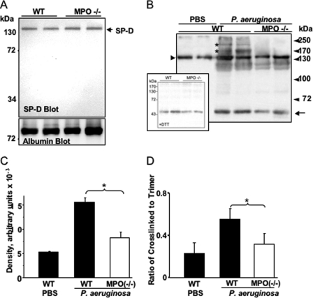
Surfactant protein D (SP-D) plays diverse and important roles in innate immunity and pulmonary homeostasis. Neutrophils and myeloperoxidase (MPO) colocalized with SP-D in a murine bacterial pneumonia model of acute inflammation, suggesting that MPO-derived reactive species might alter the function of SP-D. Exposure of SP-D to the complete MPO-H(2)O(2)-halide system caused loss of SP-D-dependent aggregating activity. Hypochlorous acid (HOCl), the major oxidant generated by MPO, caused a similar loss of aggregating activity, which was accompanied by the generation of abnormal disulfide-cross-linked oligomers. A full-length SP-D mutant lacking N-terminal cysteine residues and truncation mutants lacking the N-terminal domains were resistant to the oxidant-induced alterations in disulfide bonding. Mass spectroscopy of HOCl-treated human SP-D demonstrated several modifications, but none involved key ligand binding residues. There was detectable oxidation of cysteine 15, but no HOCl-induced cysteine modifications were observed in the C-terminal lectin domain. Together, the findings localize abnormal disulfide cross-links to the N-terminal domain. MPO-deficient mice showed decreased cross-linking of SP-D and increased SP-D-dependent aggregating activity in the pneumonia model. Thus, MPO-derived oxidants can lead to modifications of SP-D structure with associated alterations in its characteristic aggregating activity.


Figures

FIGURE 1.
FIGURE 1.
SP-D and MPO co-localize in lungs of infected mice. Mice were challenged by intranasal instillation of P. aeruginosa as described under “Experimental Procedures.” Tissues obtained 18 h after instillation of bacteria or saline were processed for immunohistochemistry. A, SP-D immunostaining of control lung using rabbit anti-mouse SP-D. B–D, representative serial sections of acutely inflamed mouse lung reacted with anti-mouse SP-D (B), antibody to MPO (C), and preimmune serum (D). SP-D specifically co-localized with MPO at sites of neutrophil infiltration. ctrl, control.
FIGURE 2.
FIGURE 2.
“MPO system” and hypochlorous acid (HOCl), but not H2O2, inhibit SP-D-dependent aggregation. A, sedimentation assays of bacterial aggregation were performed after incubation of purified rat SP-D dodecamers (2.5 μg) with the indicated components for 5 min at 37 °C in the presence of PBS containing 10 mm calcium. All assays used the same strain of K. pneumoniae as a ligand. Top, MPO system (MPO + H2O2 + NaCl). Open circles, SP-D alone; triangles, 100 nm MPO; open squares, 1 mm MPO. The MPO system significantly decreased bacterial aggregation (*, p = 0.035 for MPO at 1 μm). Diamonds, bacteria alone. Middle, hydrogen peroxide (H2O2). Open circles, SP-D alone; triangles, 1 μm H2O2; open squares, 10 mm H2O2; diamonds, bacteria alone. Bottom, hypochlorous acid. Open circles, SP-D alone; triangles, 100 μm HOCl; open squares, 1 mm; diamonds, bacteria alone. 100 μm and 1 mm HOCl significantly decreased bacterial aggregation (*, p = 0.02 for HOCl at 1 mm), but there was no detectable effect of H2O2 at concentrations as high as 10 mm. These findings are illustrative of three independent experiments per condition. The mean and S.D. (error bars) are shown. B, effects on bacterial aggregation were more directly visualized by fluorescence microscopy (×1000) using SYTO 59®-labeled Klebsiella. Rat SP-D dodecamers (2.5 μg/ml) caused microscopic and macroscopic aggregation of bacteria in PBS. Aggregation was blocked by treating SP-D with 0.1 and 1 mm HOCl for 5 min at 37 °C. Note that small aggregates could be still observed in 0.1 mm treated-SP-D. There was no effect of HOCl alone. Representative light and fluorescence images and the optical density of the suspension at 60 min are indicated. The decreased absorbance reflects clearing of the suspension as aggregated bacteria sediment to the bottom of the tube. The SP-D-dependent decrease in absorbance was reproducible and significant (A values represent the mean of three experiments; *, p = 0.015 for Klebsiella + SP-D versus Klebsiella + HOCl-treated SP-D).
FIGURE 3.
FIGURE 3.
HOCl minimally inhibits lectin activity. Lectin activity was examined using LPS bead pull-down assays as described under “Experimental Procedures.” Briefly, SP-D dodecamers were exposed to 1 mm HOCl in Hepes-buffered saline in the absence or presence of the oxidant scavenger, l-methionine (L-met). The protein was then incubated with LPS beads in the absence or presence of maltose as indicated. Bound proteins were visualized by SDS-PAGE and protein staining. Nearly all of the bead binding was inhibited by maltose. Note the efficient binding of SP-D following treatment with 1 mm HOCl (lanes 4 and 8). Mr standards are at the left. Similar findings were obtained in four independent experiments.
FIGURE 4.
FIGURE 4.
MS/MS identification of methionine sulfoxide (Met295; M+16) and methionine sulfone (Met295; M+32) in SP-D exposed to HOCl. SP-D protein (0.2 mg/ml, ∼5.6 μm) was exposed to 10 mm HOCl for 5 min at 37 °C in Hepes-buffered saline. After the reaction was terminated with l-methionine, SP-D was digested with trypsin, and the peptides were analyzed with LC-ESI-MS/MS. A, note that the y5–y10 and the b8–b11 ions had gained 16 atomic mass units (B; plus one oxygen) or 32 atomic mass units (C; plus two oxygens), but the y2–y4 and b2–b7 ions were unmodified, suggesting that Met295 in this peptide had been converted to methionine sulfoxide (M+16) or methionine sulfone (M+32).
FIGURE 5.
FIGURE 5.
Quantification of oxidized methionine (A), cysteine (B), tyrosine (C), and histidine (D) residues in SP-D exposed to HOCl. SP-D (0.2 mg/ml, ∼5.6 μm) was exposed to the indicated concentrations of HOCl as described in the legend to Fig. 4. A tryptic or Glu-C digest of SP-D was analyzed by LC-ESI-MS and MS/MS. Oxidized peptides were detected and quantified using reconstructed ion chromatograms of precursor and product peptides as described under “Experimental Procedures.” Peptide sequences were confirmed using MS/MS. Results are representative of those from two independent experiments (mean ± S.D. (error bars)).
FIGURE 6.
FIGURE 6.
HOCl alters the disulfide cross-linking of native SP-D dodecamers but does not cause disulfide cross-linking of trimeric lectin domains. A, RrSP-D dodecamers (2.5 μg) were exposed to the indicated concentration of HOCl for 5 min at 37 °C in Hepes-buffered saline in the absence or presence of excess l-methionine as scavenger (L-Met). Proteins were resolved by SDS-PAGE in the absence or presence of DTT, as indicated in the figure, and then visualized by staining with Coomassie Blue. The arrowheads identify the positions of normal unreduced trimers. Abnormal disulfide-cross-linked chains are recognized as high molecular weight species that migrate more slowly than normal trimers in the absence of reduction (asterisk). Note that 0.1 or 1 mm HOCl caused cross-linking, which is sensitive to sulfhydryl reduction. This was not observed for the incubated control (0). Non-disulfide-cross-linked components are evident as more slowly migrating species in the presence of DTT. The findings with RrSP-D were confirmed in at least four independent experiments. The extent of cross-linking was greater when incubations were performed in PBS (data not shown). The positions of Mr standards are indicated at the left. B, to confirm the origin of the high molecular weight species, treated proteins were resolved by SDS-PAGE and examined by immunoblotting with anti-SP-D. Immunoreactive high molecular weight species were generated in the presence of 1 mm HOCl (1.0) but not when SP-D was incubated with HOCl in the presence of l-methionine. The position of the normal trimer is identified (arrow). The findings are illustrative of two independent experiments. C, natural human SP-D (RhSP-D) dodecamers (2.5 μg) were treated with the indicated concentration of HOCl in PBS and resolved by SDS-PAGE as above. Unreduced cross-linked species migrating more slowly than normal trimers were generated in the presence of HOCl. In other experiments, the HOCl-dependent modifications were largely abrogated by the oxidant scavenger, l-methionine (data not shown). The normal trimer is identified (arrow), and abnormal cross-linked forms are shown by asterisks. Similar results were also obtained with native recombinant human SP-D (data not shown). D, tagless, trimeric rat NCRDs were incubated in the absence or presence of 1 mm HOCl in Hepes-buffered saline as above. There was no evidence of HOCl-dependent disulfide cross-linking. However, HOCl generated minor, non-disulfide-cross-linked species (arrows) and caused a slight increase in dispersity of the monomer band. These modifications were blocked with l-methionine. The mobilities of the cross-linked components are consistent with NCRD dimers and trimers. Mr standards are at the left. The slightly lower mobility (higher apparent mass) of proteins in the presence of DTT results from unfolding of the CRD following reduction of the normal intrachain disulfide bonds (25). The findings are representative of two independent experiments. Identical results were obtained for human NCRDs.
FIGURE 7.
FIGURE 7.
Localization of abnormal interchain disulfide cross-links to N-terminal cysteine residues. The RrSP-Dser15.20 mutant, which lacks cysteine residues within the N-terminal domains, was treated with HOCl in Hepes-buffered saline in the absence or presence of l-Met as described for dodecamers. Proteins were resolved by SDS-PAGE in the absence or presence of DTT as indicated in the figure and then visualized by staining with Coomassie Blue. Treatment with HOCl resulted in the generation of minor, non-reducible cross-linked components (brackets); this was prevented by l-methionine. No major disulfide cross-linked species were observed. Mr standards are at the left. Mobilities cannot be directly compared with the wild-type dodecamer, given greater glycosylation of the mutant protein (23). Identical findings were obtained in two independent experiments.
FIGURE 8.
FIGURE 8.
Tissue alterations in the bacterial pneumonia model. Wild-type and MPO-deficient mice were challenged by intranasal instillation of a sublethal dose of P. aeruginosa in PBS, as described under “Experimental Procedures.” Control animals received an equivalent volume of PBS. Lavage and tissues were collected at the indicated times postchallenge and processed as described. A, total BAL cells as a function of time following bacterial instillation. B, soluble peroxidase activity in cell-free BAL as a function of time following instillation. The experiments were repeated three times. Error bars, S.D. C, proteins in equivalent aliquots of cell-free BAL were resolved by SDS-PAGE and examined by immunoblotting with antibodies to MPO (top) or albumin (bottom). Elastase activity was assessed by zymography (middle), as described under “Experimental Procedures.”
FIGURE 9.
FIGURE 9.
MPO deficiency is associated with alterations in SP-D cross-linking in the setting of acute inflammation. Proteins in equivalent aliquots of cell-free BAL were resolved by SDS-PAGE and examined by immunoblotting with antibody to SP-D. A, control and MPO−/− mice show essentially identical patterns of SP-D immunostaining. B, in this representative experiment, proteins in equivalent aliquots of lavage from uninfected PBS-treated wild-type mice and infected wild-type or MPO-deficient mice were resolved by SDS-PAGE in the absence of reduction and blotted with anti-SP-D. The position of the normal SP-D trimer is indicated on the SP-D immunoblots (short arrow). Note the cross-linked species found in the lavage of wild-type animals challenged with bacteria (asterisks). The lowest molecular weight component identified in the SP-D blots (long arrow) migrates near SP-D monomers. Inset, in preliminary experiments, samples of lavage from infected wild-type and MPO-deficient mice were reduced with dithiothreitol, resolved by SDS-PAGE, and examined by immunoblotting for SP-D. C, immunoreactive components migrating between normal disulfide-cross-linked SP-D trimers and the top of the gel (B) were assessed by densitometry. The data represent the mean and S.D. of four independent assays. Cross-linked components were significantly increased in infected wild-type mice as compared with controls (p = 0.003), and cross-linked components were significantly decreased in infected MPO-deficient mice as compared with infected wild type (*, p = 0.005). D, ratios of abnormal cross-linked components over normal disulfide cross-linked trimers of SP-D for each genotype. The data represent the mean and S.D. (error bars) of four independent experiments. The ratio decreased by 2-fold in lavage fluids of MPO-deficient mice (*, p < 0.005).
FIGURE 10.
FIGURE 10.
MPO alters SP-D aggregating activity in the setting of acute inflammation. The aggregating activity of cell-free BAL was assessed by fluorescence microscopy (×200) as described above. Control experiments included bacteria alone or BAL treated with maltose before the addition of bacteria. Upper panels, representative micrographs of SYTO 59-labeled bacteria incubated with BAL from control mice (PBS-WT and PBS-MPO−/−). Middle panels, representative micrographs of SYTO 59 labeled bacteria incubated with BAL from infected WT and MPO−/− mice (infected WT and infected MPO−/−). Left lower panel, the activity of BAL from infected MPO−/− mice was examined in the presence of maltose (+Mal), the prototypical SP-D competitor. Right lower panel, the surface area of bacterial clumps formed under the above conditions was determined as described under “Experimental Procedures.” SP-D-dependent formation of bacterial clumps by BAL was reproducible. Infected MPO-deficient mice showed significantly greater bacterial aggregating activity than infected wild-type mice (gray and white bars; *, p < 0.05), which was inhibited by maltose (+Mal). Error bars, S.D. Of note, there was no significant difference in the aggregating activity of non-infected WT and MPO−/− mice (black bar) (data not shown). These findings are illustrative of four independent experiments.

Similar articles

Cited by

References

    1. Wright J. R. (2005) Nat. Rev. Immunol. 5, 58–68 - PubMed
    1. Hawgood S., Poulain F. R. (2001) Annu. Rev. Physiol. 63, 495–519 - PubMed
    1. LeVine A. M., Whitsett J. A. (2001) Microbes Infect. 3, 161–166 - PubMed
    1. Madsen J., Kliem A., Tornoe I., Skjodt K., Koch C., Holmskov U. (2000) J. Immunol. 164, 5866–5870 - PubMed
    1. Crouch E. C. (2000) Respir. Res. 1, 93–108 - PMC - PubMed

Publication types