. 2010 May 14;285(20):14909-14919.
doi: 10.1074/jbc.M110.106724. Epub 2010 Mar 12.

Role of protein misfolding in DFNA9 hearing loss

Affiliations

Role of protein misfolding in DFNA9 hearing loss

Jianhua Yao et al. J Biol Chem. .

Abstract

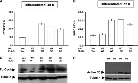
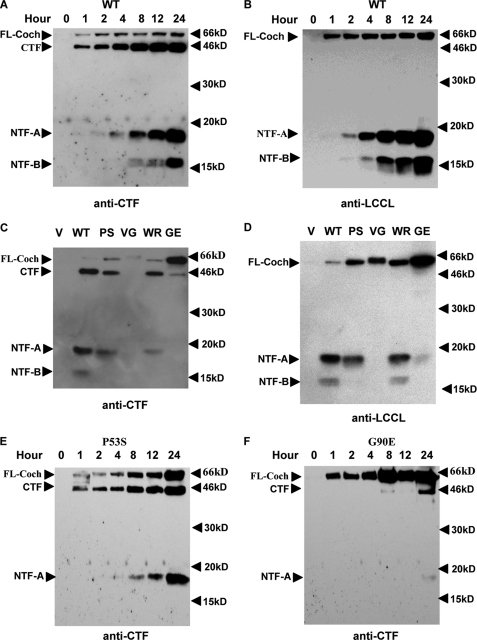
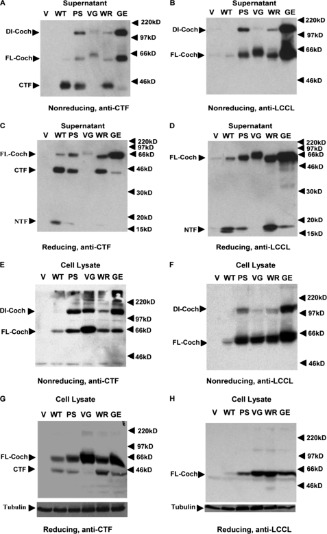
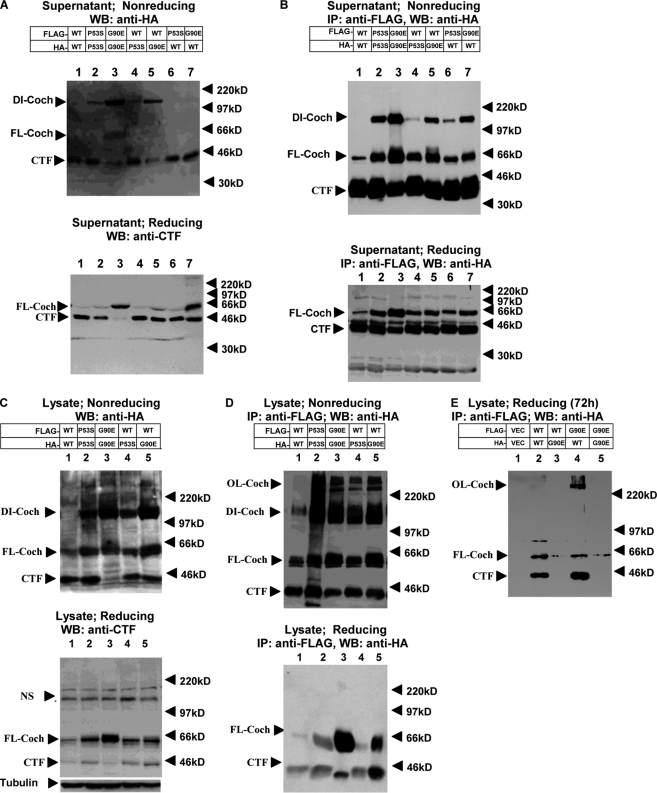
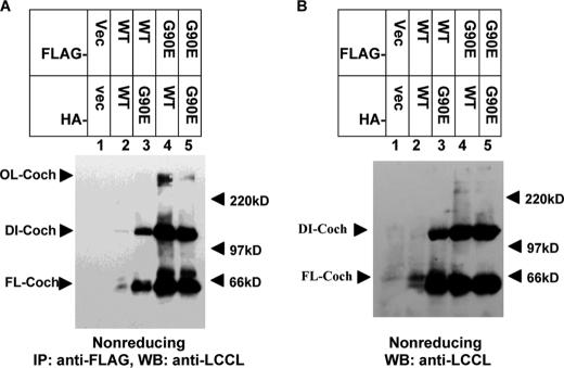
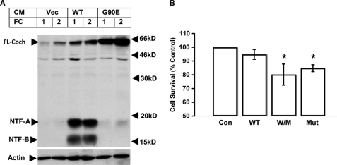
Mutations in the COCH (coagulation factor C homology) gene have been attributed to DFNA9 (deafness, autosomal-dominant 9), an autosomal-dominant non-syndromic hearing loss disorder. However, the mechanisms responsible for DFNA9 hearing loss remain unknown. Here, we demonstrate that mutant cochlin, the protein product of the COCH gene, forms a stable dimer that is sensitive to reducing agent. In contrast, wild-type (WT) cochlin may form only dimers transiently. Interestingly, the presence of mutant cochlin can stabilize WT cochlin in dimer conformation, providing a possible mechanism for the dominant nature of DFNA9 mutations. Furthermore, the expression of mutant cochlin eventually induces WT cochlin to form stable oligomers that are resistant to reducing agent. Finally, we show that mutant cochlin is cytotoxic in vitro and in vivo. Our study suggests a possible molecular mechanism underlying DFNA9 hearing loss and provides an in vitro model that may be used to explore protein-misfolding diseases in general.


Figures

FIGURE 1.
FIGURE 1.
Cell death induced by coexpressing WT and mutant cochlins. UB/UE-1 cells were cotransfected with HA- and FLAG-tagged WT and mutant G90E (GE) cochlins as indicated. Cell death was measured after UB/UE-1 cells were transfected for 48 h (A) or 72 h (B) by counting green fluorescent protein-positive live versus dead cells. At least 200 cells were counted for each of treatment, and at least three independent experiments were performed. Total cochlin expression was determined by Western blotting using anti-LCCL monoclonal antibody against cochlin (C). Activation of caspase-3 was detected by Western blotting using anti-active caspase-3 antibody (D). Student's t test was applied for statistical analysis. Vec, vector; FL-Coch, full-length cochlin; PS, P53S.
FIGURE 2.
FIGURE 2.
Dynamics of cochlin secretion. WT (A and B) and mutant (C–F) cochlin expression vectors were transiently transfected into HEK293T cells, and the culture supernatant was collected at the indicated time points (A, B, E, and F) and analyzed by Western blotting using anti-CTF polyclonal antibody (A, C, E, and F) or anti-LCCL monoclonal antibody (B and D) after separation by 15% reducing SDS-PAGE. The results shown are representative of three separate experiments. FL-Coch, full-length cochlin; V, vector; PS, P53S; VG, V68G; WR, W119R; GE, G90E.
FIGURE 3.
FIGURE 3.
COCH mutations induce the formation of a 120-kDa mutant complex intracellularly and extracellularly. The culture supernatant (A–D) or cell lysates (E–H) from transiently transfected HEK293T cells expressing the control vector (V), the WT COCH gene, or mutant P53S (PS), V68G (VG), G90E (GE), or W119R (WR) COCH were separated by 8% nonreducing SDS-PAGE (A, B, E, and F) or by 12% (C and D) or 10% (G and H) reducing SDS-PAGE. The Western blots were probed with anti-CTF polyclonal antibody (A, C, E, and G) or anti-LCCL monoclonal antibody (B, D, F, and H). The arrowheads point to the 120-kDa mutant-specific band (dimer cochlin (DI-Coch)), full-length cochlin (FL-Coch), CTF, and NTF.
FIGURE 4.
FIGURE 4.
Dimerization of cochlins. HEK293T cells were cotransfected with HA- or FLAG-tagged WT, P53S, or G90E cochlin as indicated at a 1:1 ratio. Forty-eight hours after transfection (A–D), supernatants and lysates were collected. Western blotting (WB) of the culture supernatant (A and B) and cell lysates (C and D) was performed using anti-HA antibody after separation by 8% nonreducing SDS-PAGE (A and C, upper panels) or anti-CTF antibody after separation by 12% reducing SDS-PAGE (A and C, lower panels). The collected culture supernatant and lysates (B and D) were used to perform immunoprecipitation (IP) with anti-FLAG antibody-conjugated agarose beads. After washing, the FLAG immunocomplexes were eluted with FLAG peptide, and the eluates were analyzed by 8% nonreducing SDS-PAGE (B and D, upper panels) or 12% reducing SDS-PAGE (B and D, lower panels). The Western blots were probed with anti-HA antibody. The bands at ∼220 kDa in C (both upper and lower panels) are nonspecific. NS, nonspecific band. Cell lysates were collected 72 h after transfection, and 12% SDS-PAGE was conducted under reducing conditions (E). The Western blots were probed with anti-HA antibody. The results shown are representative of three independent experiments. DI-Coch, dimer cochlin; FL-Coch, full-length cochlin; OL-Coch, oligomer cochlin.
FIGURE 5.
FIGURE 5.
WT and mutant cochlin oligomerization in UB/UE-1 cells. UB/UE-1 cells were cotransfected with HA- and FLAG-tagged WT and mutant G90E cochlins as indicated at a 1:1 ratio. Cochlin was detected by immunoprecipitation (IP) using anti-FLAG antibody, and the Western blot (WB) was probed with anti-LCCL antibody after 8% nonreducing SDS-PAGE (A). Also shown is a control Western blot probed with anti-LCCL antibody after nonreducing SDS-PAGE of the cell lysates shown in A (B). Vec, vector; DI-Coch, dimer cochlin; OL-Coch, oligomer cochlin; FL-Coch, full-length cochlin.
FIGURE 6.
FIGURE 6.
Mutant cochlin induces hearing loss and histological changes in the cochlea. WT and mutant G90E (Mut) cochlins were transiently expressed in HEK293T cells. Special culture medium without fetal bovine serum and antibiotics (CM) was collected and concentrated and then injected into the cochleae of adult C57BL/6 mice (n = 4), and the ABR thresholds were measured 1 and 4 weeks after injection (A). The thickness of the stria vascularis (Stv) was measured in hematoxylin/eosin-stained mouse inner ears (B). The morphological changes in mouse cochleae injected with WT CM (C) and mutant G90E cochlin (D) collected at 4 weeks after injection are shown (*, p < 0.05, t test), with arrows pointing to the stria vascularis. Four different types of fibrocytes affected by injection of G90E CM are clearly observed in the spiral ligament (SPL) but not in the spiral limbus (E).
FIGURE 7.
FIGURE 7.
Extracellular mutant cochlin binds to primary fibrocytes and reduces cell survival. A, fibrocytes (FC) from WT mice were isolated and cultured in vitro and treated with CM (1:5 dilution) collected from transiently transfected HEK293T cells expressing vehicle control (Vec) or WT or mutant G90E cochlin. After treatment, the supernatant was removed, and cells were washed twice with phosphate-buffered saline. The cell lysates were analyzed by 15% reducing SDS-PAGE, followed by Western blotting using anti-LCCL antibody. B, primary fibrocytes were incubated with CM from 293T cells expressing vehicle (Con), WT cochlin alone, WT and mutant G90E cochlins together (W/M), or mutant G90E cochlin alone (Mut) for 72 h. Cell survival was measured using the MTS assay. *, p < 0.05. FL-Coch, full-length cochlin.

References

    1. Bossy-Wetzel E., Schwarzenbacher R., Lipton S. A. (2004) Nat. Med. 10, (suppl.) S2–S9 - PubMed
    1. Robertson N. G., Lu L., Heller S., Merchant S. N., Eavey R. D., McKenna M., Nadol J. B., Jr., Miyamoto R. T., Linthicum F. H., Jr., Lubianca Neto J. F., Hudspeth A. J., Seidman C. E., Morton C. C., Seidman J. G. (1998) Nat. Genet. 20, 299–303 - PubMed
    1. Merchant S. N., Linthicum F. H., Nadol J. B., Jr. (2000) Adv. Otorhinolaryngol. 56, 212–217 - PubMed
    1. Khetarpal U., Schuknecht H. F., Gacek R. R., Holmes L. B. (1991) Arch. Otolaryngol. Head Neck Surg. 117, 1032–1042 - PubMed
    1. Robertson N. G., Skvorak A. B., Yin Y., Weremowicz S., Johnson K. R., Kovatch K. A., Battey J. F., Bieber F. R., Morton C. C. (1997) Genomics 46, 345–354 - PubMed

Publication types