. 2010 Jun;35(5):351-61.
doi: 10.1093/chemse/bjq019. Epub 2010 Mar 12.

Rapid degeneration and regeneration of the zebrafish olfactory epithelium after triton X-100 application

Affiliations

Rapid degeneration and regeneration of the zebrafish olfactory epithelium after triton X-100 application

Tania Iqbal et al. Chem Senses. 2010 Jun.

Abstract

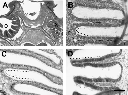
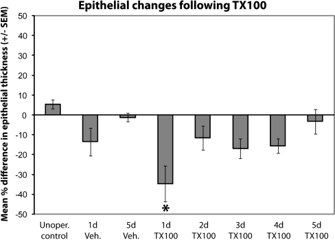
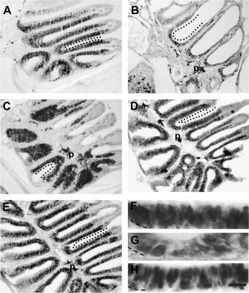
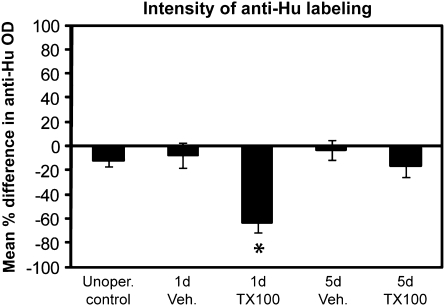
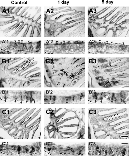
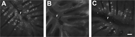
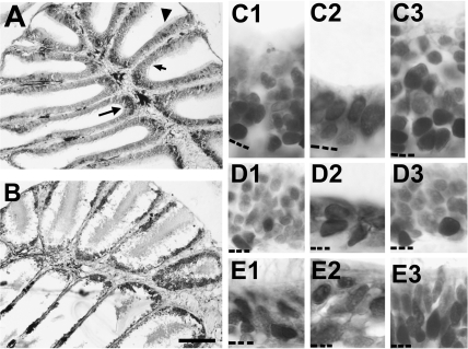
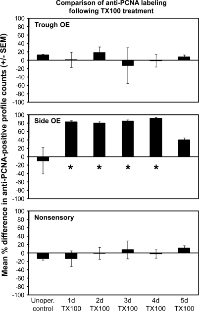
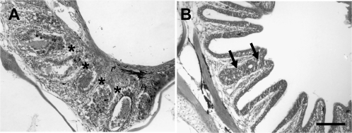
The effects of Triton X-100 on the olfactory epithelium (OE) of adult zebrafish were examined to study neuronal turnover in this model system. Fish were killed at various time points after detergent application and stained with hematoxylin and eosin to examine olfactory structures, immunocytochemistry to examine cell types, or DiI to examine connections to the olfactory bulb. A significant decrease in epithelial thickness of treated sides was observed 1-day posttreatment. Epithelium thickness recovered by 5 days. The most significant reduction in the OE following Triton X-100 treatment corresponded to the region of supporting cells and mature olfactory sensory neurons. Labeling for all neurons with anti-Hu and for the 3 sensory neuron subtypes of the zebrafish OE (ciliated, microvillous, and crypt neurons) diminished 1 day after lesion and returned by 5 days posttreatment. Retrograde labeling from the olfactory bulb showed that the majority of mature olfactory sensory neurons disappeared in 1 day and reappeared by 5 days after treatment. Anti-proliferating cell nuclear antigen was used to show mitotic activity, and after chemical lesion, there was an increase in proliferation in specific regions of the OE. Thus, chemical ablation causes temporary reduction with swift regeneration of the OE occurring within a week.


Figures

Figure 1
Figure 1
Analysis of hematoxylin and eosin-stained horizontal sections through the whole head showed obvious morphological changes in the OE following Triton X-100 treatment. (A) Immediately after Triton X-100 was applied to the olfactory organ, the integrity of the right OE (x) was drastically compromised, with cellular debris visible throughout the nasal cavity. The left olfactory organ was not affected (o), and the multiple lamellae that make up the rosette were present and intact. (B) Vehicle-treated olfactory organs showed no alterations in lamellar morphology or epithelial structure 1 day after application. An example of an area of sensory epithelium adjacent to the central raphe (r) is shown with a dotted line. (C) One day following Triton X-100 application, the depth from the apical surface to the basal membrane of the OE appeared reduced. A dotted line marks an area of OE, which includes troughs adjacent to the central raphe (r) and the medial portion of lamellae. (D) By 5 days posttreatment, the epithelium no longer appeared thinned. The sensory epithelium, indicated by a dotted line, is found in this section on the portion of lamellae medial to the central raphe (to the left in the micrograph but not present in this section). Scale bar = 200 μm for A, 50 μm for BD.
Figure 2
Figure 2
Quantitative analysis of olfactory epithelial thickness following Triton X-100 application allowed comparison of epithelial changes. The percent difference in thickness of the OE between the internal control and treated sides of the 1 day post-detergent application group was confirmed by ANOVA to be different from unoperated control, 5 days post-vehicle, and 5 days post-lesion groups. For each group n = 3 and P < 0.05 was considered significant.
Figure 3
Figure 3
Examination of distribution of mature and immature neurons using α-Hu analysis. (A) Heavy labeling for α-Hu was present in the sensory regions of the control rosette. (B) One day following chemical ablation of the OE, there were few α-Hu immunoreactive profiles in the olfactory rosette. (C) By 5 days after Triton X-100 treatment, the amount of α-Hu labeling had increased dramatically throughout the sensory areas of the rosette. Seven (D) and 14 (E) days after detergent treatment, Hu-positive profiles populated the entire OE. Control (F), 1d (G), and 5d (H) α-Hu labeled OEs at higher magnification show cellular distribution of the antibody label. Basement membrane is marked with a dashed line. Demarcated with dotted line (AE) are areas of sensory epithelium along lamellae. p = pigment of the lamina propria in the central raphe and is not indicative of antibody labeling. Scale bar = 100 μm for AE and 10 μm for FH.
Figure 4
Figure 4
Quantitative analysis of α-Hu OD allowed evaluation of changes to numbers of neurons present in epithelium. Percent differences in OD levels between treated and untreated nares of 3 animals from each group were calculated, and ANOVA was employed to confirm differences (P < 0.05 was considered significant). There was a significant difference in OD 1 day post Triton X-100 compared with unoperated controls and 5 day groups.
Figure 5
Figure 5
Antibodies were used to characterize the effects of chemical ablation on specific cell types of the zebrafish OE. (A) Anti-Gα s/olf in control epithelia (A1) revealed immunoreactive profiles throughout the sensory region of the rosette. Dendritic knobs and cilia were distinctly labeled (A′1, arrowheads), and there was staining found diffusely through the depth of the epithelium except the basal region. One day after Triton X-100 application, there were fewer Gα s/olf -positive areas in the epithelium (A2), but where labeling occurred, the dendritic knobs and cilia of ciliated neurons were marked (A′2) with only faint labeling of the epithelium underneath. Five days after chemical lesion (A3), however, α-Gα s/olf staining had returned to normal levels and pattern (A′3). (B) Anti-calretinin labeling in control olfactory organs identified discrete cell bodies and their dendritic processes (B′1, arrows) scattered throughout the OE (B1). Immunoreactivity to this cell marker was greatly diminished 1 day following Triton X-100 treatment (B2), and those cells labeled appeared to be without dendritic knobs (B′2). Labeling to anti-calretinin was increased by 5 days after treatment (B3), and the bodies that were labeled also had obvious processes (B′3). (C) Anti-S100 labeled large round cells (^ in C′1), the crypt neurons, scattered around the sensory regions in the OE of control rosettes (C1). S100-immunoreactivity showed an overall decrease 1 day after treatment (C2), and the bodies were smaller and flattened compared with control (C′2). Immunoreactivity recovered to near control levels (C3) and morphology (C′3) 5 days after lesioning. Demarcated with dotted lines are examples of areas where labeling was present within the sensory epithelium. p = pigment of the lamina propria in the central raphe and is not indicative of antibody labeling. Basement membrane is marked with a dashed line. Scale bar = 100 μm for A1–3, B1–3, C1–3 and 10 μm for A′1–3, B′1–3, C′1–3.
Figure 6
Figure 6
Retrograde labeling from the olfactory bulb using DiI showed numerous mature olfactory sensory neurons throughout the rosette of control animals (A). There was little or no DiI labeling in the rosettes of animals examined 1 day after Triton X-100 treatment (B), but there were numerous DiI-labeled cells 5 days after detergent application (C). r = central raphe. Scale bar = 50 μm for all.
Figure 7
Figure 7
Anti-PCNA-immunoreactivity was observed in unoperated animals (A) in the basal region of the OE throughout the rosette, with higher levels of staining in the troughs between lamellae (longer arrow) as well as in the nonsensory regions (arrowhead). The side OE on the medial extent of the lamellae (shorter arrow) had less labeling. (B) Immediately after Triton X-100 application, significant damage to the apical regions of the epithelium throughout the rosette was apparent. Almost all the remaining cells were anti-PCNA positive. Higher magnification images of the trough OE (C), side OE (D), and nonsensory (E) regions of unoperated controls (C1, D1, E1), 1-day treated (C2, D2, E2), and 5-day treated (C3, D3, E3) animals revealed different patterns of cell proliferation. Control animals had numerous PCNA-positive profiles throughout the trough region (C1), few in the side OE region (D1), and many in the nonsensory region (E1). One day after detergent treatment, the epithelium was thinner, but the remaining cells in all 3 regions were heavily labeled for anti-PCNA (C2, D2, E2). Five days after detergent application, epithelial thickness returned and the pattern of antibody labeling was similar to controls (C3, D3, E3). Basement membrane is marked with a dashed line. Scale bar = 100 μm for A, B and 10 μm for C1–E3.
Figure 8
Figure 8
Quantitative analysis of anti-PCNA allowed comparison of mitotic activity in unoperated animals and animals allowed to survive for various time periods following Triton X-100 treatment. The percent differences in average number of anti-PCNA labeled profiles in a 25 × 50 μm region of the trough OE, side OE, and nonsensory regions of right and left olfactory organs were compared with ANOVA. There was a significant difference in the side OE region at 1d, 2d, 3d, and 4d postdetergent application. For each group n = 3 and P < 0.05 was considered significant.
Figure 9
Figure 9
(A) Some additional morphological abnormalities were sometimes observed following Triton X-100 treatment. At survival times of 1 or 2 days, changes to the structure of the olfactory rosette were observed occasionally, such as complete fusion of lamellae. Although lamellar morphology was destroyed, remnants of olfactory tissue were still visible (asterisks). (B) Sometimes seen at later time points was fusion between 2 adjacent lamellae (arrows). Scale bar = 100 μm for both.

Similar articles

Cited by

References

    1. Adamek GD, Gesteland RC, Mair RG, Oakley B. Transduction physiology of olfactory receptor cilia. Brain Res. 1984;310:87–97. - PubMed
    1. Adolf B, Chapouton P, Lam CS, Topp S. Conserved and acquired features of adult neurogenesis in the zebrafish telencephalon. Dev Biol. 2006;295:278–293. - PubMed
    1. Baker H, Kawano T, Margolis FL, Joh TH. Transneuronal regulation of tyrosine hydroxylase expression in olfactory bulb of mouse and rat. J Neurosci. 1983;3:69–78. - PMC - PubMed
    1. Barth AL, Justice NJ, Ngai J. Asynchronous onset of odorant receptor expression in the developing zebrafish olfactory system. Neuron. 1996;16:23–34. - PubMed
    1. Becker CG, Becker T. Adult zebrafish as a model for successful central nervous system regeneration. Restor Neurol Neurosci. 2008;26:71–80. - PubMed

Publication types

LinkOut - more resources