

CXCR3/ligands are significantly involved in the tumorigenesis of basal cell carcinomas
- PMID: 20228225
- PMCID: PMC2861108
- DOI: 10.2353/ajpath.2010.081059
CXCR3/ligands are significantly involved in the tumorigenesis of basal cell carcinomas
Abstract
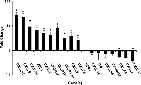
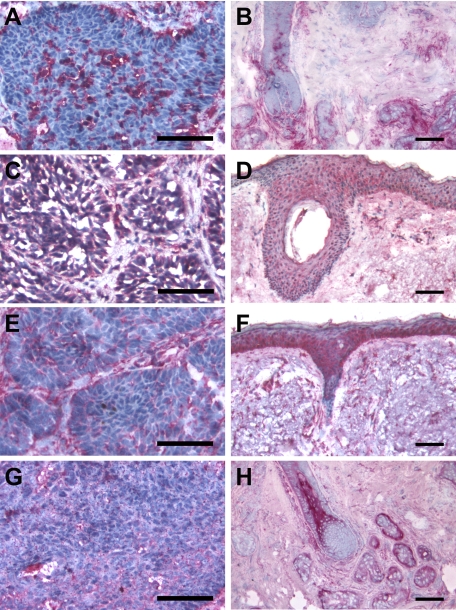
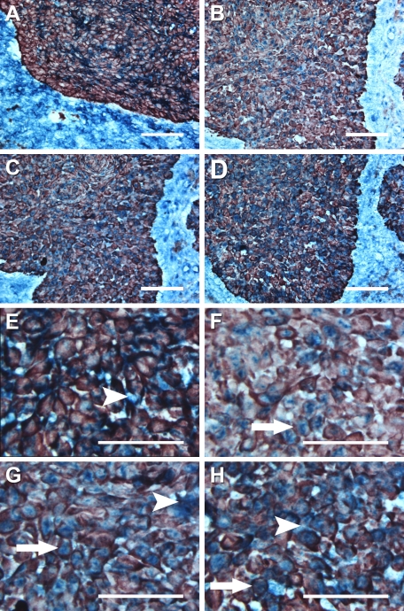
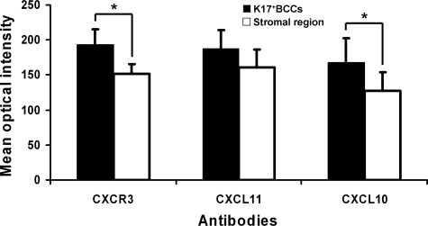
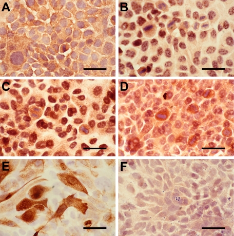
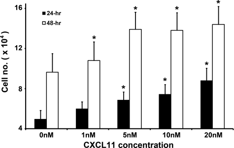
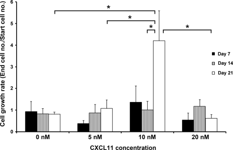
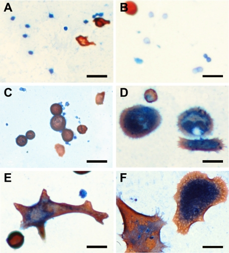
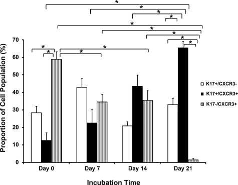
Basal cell carcinoma (BCC) is the most common skin malignancy encountered worldwide. We hypothesized that CXC chemokines, small cytokines involved in inducing directed leukocyte chemotaxis, could play a key role in the modulation of BCC growth. In this study, quantitative RT-PCR revealed that the chemokines CXCL9, 10, 11, and their receptor CXCR3 were significantly upregulated by an average 22.6-fold, 9.2-fold, 26.6-fold, and 4.9-fold, respectively in BCC tissue samples as compared with nonlesional skin epithelium. Immunohistochemistry analysis revealed that CXCR3, CXCL10, and CXCL11, but not CXCL9, colocalized with cytokeratin 17 (K17) in BCC keratinocytes. In addition, CXCR3 and its ligands were expressed in cells of the surrounding BCC stroma. The chemokines and K17 were also expressed in cultured human immortalized HaCaT keratinocytes. Exposure of HaCaT cells or primary BCC-derived cells to CXCL11 peptides in vitro significantly increased cell proliferation. In primary BCC-derived cell cultures, addition of CXCL11 progressively selected for K17+/CXCR3+ co-expressing cells over time. The expression of CXCR3 and its ligands in human BCC keratinocytes, the enhancement of keratinocyte cell proliferation by CXCL11, and the homogeneity of K17+ BCC cells in human BCC-isolated cell population supported by CXCR3/CXCL11 signaling all suggest that CXCR3 and its ligands may be important autocrine and/or paracrine signaling mediators in the tumorigenesis of BCC.
Figures
Comment in
-
Roles for aberrant CXCR3 signaling in basal cell carcinoma: a case for dual activity.Am J Pathol. 2010 May;176(5):2088-91. doi: 10.2353/ajpath.2010.091284. Epub 2010 Feb 25. Am J Pathol. 2010. PMID: 20185576 Free PMC article.
Similar articles
-
CXCR3 ligands promote expression of functional indoleamine 2,3-dioxygenase in basal cell carcinoma keratinocytes.Br J Dermatol. 2011 Nov;165(5):1030-6. doi: 10.1111/j.1365-2133.2011.10489.x. Epub 2011 Oct 17. Br J Dermatol. 2011. PMID: 21711334
-
Regulation of C-X-C chemokine gene expression by keratin 17 and hnRNP K in skin tumor keratinocytes.J Cell Biol. 2015 Mar 2;208(5):613-27. doi: 10.1083/jcb.201408026. Epub 2015 Feb 23. J Cell Biol. 2015. PMID: 25713416 Free PMC article.
-
Modelling of the membrane receptor CXCR3 and its complexes with CXCL9, CXCL10 and CXCL11 chemokines: putative target for new drug design.Mol Immunol. 2009 Dec;47(2-3):332-9. doi: 10.1016/j.molimm.2009.09.013. Epub 2009 Oct 1. Mol Immunol. 2009. PMID: 19800124
-
Review: The chemokine receptor CXCR3 and its ligands CXCL9, CXCL10 and CXCL11 in neuroimmunity--a tale of conflict and conundrum.Neuropathol Appl Neurobiol. 2010 Aug;36(5):368-87. doi: 10.1111/j.1365-2990.2010.01089.x. Epub 2010 May 6. Neuropathol Appl Neurobiol. 2010. PMID: 20487305 Review.
-
CXCL9, CXCL10, CXCL11/CXCR3 axis for immune activation - A target for novel cancer therapy.Cancer Treat Rev. 2018 Feb;63:40-47. doi: 10.1016/j.ctrv.2017.11.007. Epub 2017 Nov 26. Cancer Treat Rev. 2018. PMID: 29207310 Free PMC article. Review.
Cited by
-
The expanding significance of keratin intermediate filaments in normal and diseased epithelia.Curr Opin Cell Biol. 2013 Feb;25(1):47-56. doi: 10.1016/j.ceb.2012.10.018. Epub 2012 Dec 25. Curr Opin Cell Biol. 2013. PMID: 23270662 Free PMC article. Review.
-
Potential Role of CXCL10 in Monitoring Response to Treatment in Leprosy Patients.Front Immunol. 2021 Jul 20;12:662307. doi: 10.3389/fimmu.2021.662307. eCollection 2021. Front Immunol. 2021. PMID: 34354699 Free PMC article.
-
Outcome markers of ART-treated HIV+ patients with early stage Kaposi's sarcoma.PLoS One. 2020 Jul 7;15(7):e0235865. doi: 10.1371/journal.pone.0235865. eCollection 2020. PLoS One. 2020. PMID: 32634155 Free PMC article.
-
Keratin intermediate filament proteins - novel regulators of inflammation and immunity in skin.J Cell Sci. 2012 Nov 15;125(Pt 22):5257-8. doi: 10.1242/jcs.122929. J Cell Sci. 2012. PMID: 23377656 Free PMC article. No abstract available.
-
Kynurenic Acid and Its Analog SZR104 Exhibit Strong Antiinflammatory Effects and Alter the Intracellular Distribution and Methylation Patterns of H3 Histones in Immunochallenged Microglia-Enriched Cultures of Newborn Rat Brains.Int J Mol Sci. 2022 Jan 19;23(3):1079. doi: 10.3390/ijms23031079. Int J Mol Sci. 2022. PMID: 35163002 Free PMC article.
References
-
- Aszterbaum M, Epstein J, Oro A, Douglas V, LeBoit PE, Scott MP, Epstein EH., Jr Ultraviolet and ionizing radiation enhance the growth of BCCs and trichoblastomas in patched heterozygous knockout mice. Nat Med. 1999;5:1285–1291. - PubMed
-
- Walling HW, Fosko SW, Geraminejad PA, Whitaker DC, Arpey CJ. Aggressive basal cell carcinoma: presentation, pathogenesis, and management. Cancer Metastasis Rev. 2004;23:389–402. - PubMed
-
- Diepgen TL, Mahler V. The epidemiology of skin cancer. Br J Dermatol. 2002;146 Suppl 61:1–6. - PubMed
-
- Gallagher RP, Ma B, McLean DI, Yang CP, Ho V, Carruthers JA, Warshawski LM. Trends in basal cell carcinoma, squamous cell carcinoma, and melanoma of the skin from 1973 through 1987. J Am Acad Dermatol. 1990;23:413–421. - PubMed
-
- Marks R, Staples M, Giles GG. Trends in non-melanocytic skin cancer treated in Australia: the second national survey. Int J Cancer. 1993;53:585–590. - PubMed
Publication types
MeSH terms
Substances
Grants and funding
LinkOut - more resources
Full Text Sources
Other Literature Sources
Medical
Molecular Biology Databases
Research Materials
