

Sustained VEGF delivery via PLGA nanoparticles promotes vascular growth
- PMID: 20228260
- PMCID: PMC2886627
- DOI: 10.1152/ajpheart.00199.2009
Sustained VEGF delivery via PLGA nanoparticles promotes vascular growth
Abstract
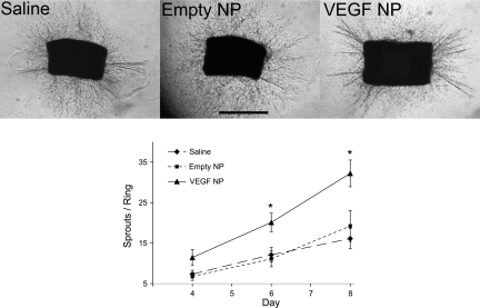
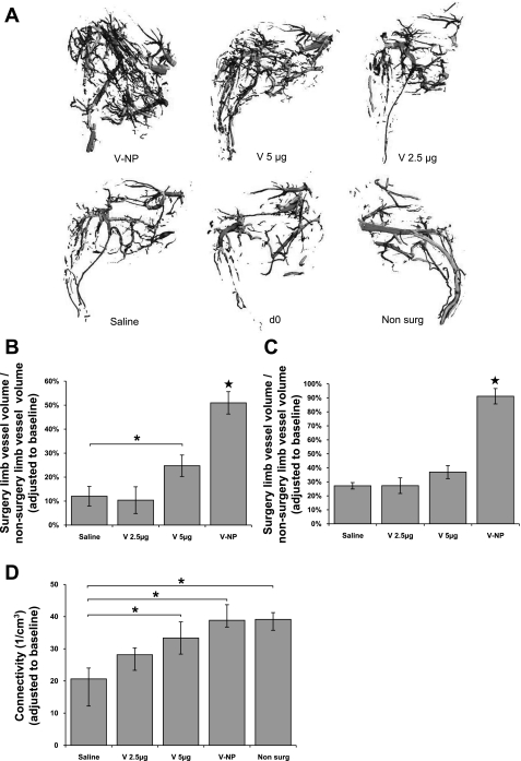
Technologies to increase tissue vascularity are critically important to the fields of tissue engineering and cardiovascular medicine. Currently, limited technologies exist to encourage angiogenesis and arteriogenesis in a controlled manner. In the present study, we describe an injectable controlled release system consisting of VEGF encapsulated in poly(lactic-co-glycolic acid) (PLGA) nanoparticles (NPs). The majority of VEGF was released gradually over 2-4 days from the NPs as determined by an ELISA release kinetics experiment. An in vitro aortic ring bioassay was used to verify the bioactivity of VEGF-NPs compared with empty NPs and no treatment. A mouse femoral artery ischemia model was then used to measure revascularization in VEGF-NP-treated limbs compared with limbs treated with naked VEGF and saline. 129/Sv mice were anesthetized with isoflurane, and a region of the common femoral artery and vein was ligated and excised. Mice were then injected with VEGF-NPs, naked VEGF, or saline. After 4 days, three-dimensional microcomputed tomography angiography was used to quantify vessel growth and morphology. Mice that received VEGF-NP treatment showed a significant increase in total vessel volume and vessel connectivity compared with 5 microg VEGF, 2.5 microg VEGF, and saline treatment (all P < 0.001). When the yield of the fabrication process was taken into account, VEGF-NPs were over an order of magnitude more potent than naked VEGF in increasing blood vessel volume. Differences between the VEGF-NP group and all other groups were even greater when only small-sized vessels under 300 mum diameter were analyzed. In conclusion, sustained VEGF delivery via PLGA NPs shows promise for encouraging blood vessel growth in tissue engineering and cardiovascular medicine applications.
Figures


References
-
- Davda J, Labhasetwar V. Characterization of nanoparticle uptake by endothelial cells. Int J Pharm 233: 51–59, 2002 - PubMed
-
- Duvall CL, Taylor WR, Weiss D, Guldberg RE. Quantitative microcomputed tomography analysis of collateral vessel development after ischemic injury. Am J Physiol Heart Circ Physiol 287: H302–H310, 2004 - PubMed
-
- Duvall CL, Weiss D, Robinson ST, Alameddine FM, Guldberg RE, Taylor WR. The role of osteopontin in recovery from hind limb ischemia. Arterioscler Thromb Vasc Biol 28: 290–295, 2008 - PubMed
-
- Freedman SB, Isner JM. Therapeutic angiogenesis for coronary artery disease. Ann Intern Med 136: 54–71, 2002 - PubMed
Publication types
MeSH terms
Substances
Grants and funding
LinkOut - more resources
Full Text Sources
Other Literature Sources
Miscellaneous