. 2010 Apr;213(Pt 7):1182-94.
doi: 10.1242/jeb.040378.

Octopamine promotes rhythmicity but not synchrony in a bilateral pair of bursting motor neurons in the feeding circuit of Aplysia

Affiliations

Octopamine promotes rhythmicity but not synchrony in a bilateral pair of bursting motor neurons in the feeding circuit of Aplysia

C Martínez-Rubio et al. J Exp Biol. 2010 Apr.

Abstract

Octopamine-like immunoreactivity was localized to a limited number (<40) of neurons in the Aplysia central nervous system, including three neurons in the paired buccal ganglia (BG) that control feeding movements. Application of octopamine (OA) to the BG circuit produced concentration-dependent (10(-8)-10(-4) mol l(-1)) modulatory actions on the spontaneous burst activity of the bilaterally paired B67 pharyngeal motor neurons (MNs). OA increased B67's burst duration and the number of impulses per burst. These effects reflected actions of OA on the intrinsic tetrodotoxin-resistant driver potential (DP) that underlies B67 bursting. In addition to its effects on B67's burst parameters, OA also increased the rate and regularity of burst timing. Although the bilaterally paired B67 MNs both exhibited rhythmic bursting in the presence of OA, they did not become synchronized. In this respect, the response to OA differed from that of dopamine, another modulator of the feeding motor network, which produces both rhythmicity and synchrony of bursting in the paired B67 neurons. It is proposed that modulators can regulate burst synchrony of MNs by exerting a dual control over their intrinsic rhythmicity and their reciprocal capacity to generate membrane potential perturbations. In this simple system, dopaminergic and octopaminergic modulation could influence whether pharyngeal contractions occur in a bilaterally synchronous or asynchronous fashion.


Figures
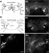
Fig. 1.
Fig. 1.
Octopamine-like immunoreactivity (OAli) in the buccal and cerebral ganglia. (Ai) Schematic summary of OAli in the paired buccal ganglia. Strong OAli was limited to three moderately sized neurons. Broken rectangle indicates region shown in Aii. Abbreviations: s n., salivary nerve; rad n., radula nerve; e n., esophageal nerve; bn1, buccal nerve 1; bn2, buccal nerve 2; bn3, buccal nerve 3; Cb c., cerebral-buccal connective. (Aii) Two of the OAli neurons (arrows) were positioned symmetrically in the medial region of the caudal surface of each hemiganglion. These cells gave rise to fibers that traversed the buccal commissure (b c.). An unpaired cell (arrowhead) was located within the b c. Calibration bar=100 μm. (Bi) Schematic summary of OAli in the cerebral ganglion. Approximately six cells were symmetrically positioned on the dorsal surface of the lateral region of each hemiganglion. Broken rectangle indicates region shown in (Biii). Abbreviations: Ul n., upper labial nerve; At n., anterior tentacular nerve; Cpd c., cerebral-pedal connective; Cpl c., cerebral-pleural connective. (Bii) Low power image of the dorsal surface of the cerebral ganglion. Cluster of approximately four small (~20 μm diameter) cells was located slightly anterior to the origin of each cerebral-pedal connective (arrows). Additional individual bilateral neurons (arrowheads) are present in a more anterior position. Calibration bar= 200 μm. (Biii) Higher magnification of OAli neurons in the left cerebral hemiganglion. Two slightly larger (40–50 μm) cells were located separately (arrowheads), one anterior to the cluster and another more posterior, between the origins of the cerebral-pedal and cerebral-pleural connectives. Calibration bar=100 μm. (Biv) Upper quandrant of the ventral surface of the cerebral ganglion. No OAli neurons were detected on the ventral surface but immunoreactive fiber plexuses were observed near the origin of the cerebral-buccal connective (C-b c.) (arrow) and in the region of the M cluster (arrowhead). Calibration bar=100 μm.
Fig. 2.
Fig. 2.
Octopamine-like immunoreactivity (OAli) in the pedal ganglia. (A) Schematic drawing showing the positions of OAli neurons in the paired pedal ganglia (Ped g.). All of the pedal OAli neurons were bilaterally paired. The cells on the dorsal surface of each ganglion are shown on the left side of this drawing and the neurons on the ventral surface are shown on the right. The parapedal commissure and the unpaired parapedal commissure nerve (P10) contained several strongly staining OAli fibers. Broken rectangles mark the areas of each ganglion shown in panels B and C. Abbreviations: Pl g., pleural ganglion; C-pl c., cerebral-pleural connectives; C-p c., cerebral-pedal connective; P c., pedal commissure; Pp c., parapedal commissure. (B) Three intensely stained cell bodies (approximately 75 μm diameter) were clustered in the central region of the ventral surface of each pedal ganglion. Each soma appeared to give rise to multiple large protrusions (arrow) and fine fibers (arrowhead). (C) Smaller OAli neurons in the lateral region of the dorsal surface of each pedal ganglion. A tightly grouped cluster of approximately six small (20 μm diameter) cells was located slightly anterior to the origins of pedal nerves P6 and P7 (arrow). Two slightly larger (approximately 40 μm) cells were located separately (arrowheads), one anterior to the cluster and another more posterior, near the origin of P8. Scale bar=100 μm, applies to panels B and C.
Fig. 3.
Fig. 3.
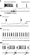
Effects of octopamine (OA) on B67 in normal artificial seawater. (A) Control recordings. Upper trace: intracellular recording from B67. Lower trace: extracellular recording from the radula nerve (Rn). B67 exhibited two modes of patterned activity in the isolated cerebral-buccal preparation. It was recruited into multi-phasic buccal motor programs (BMPs) where it fired during the phase of fictive radula protraction (shaded bar below recording) and was inhibited during retraction (white bar). B67 also exhibited a monophasic endogenous burst pattern that was previously shown to reflect the intrinsic properties of this cell (Serrano and Miller, 2006). In contrast to the spontaneous bursts, which were detected only in B67, the BMPs involved a large number of buccal neurons (compare radula nerve recording below each pattern; B67 does not project to the Rn). The BMPs occurred less frequently (<0.01 Hz) than the spontaneous intrinsic bursts (<0.1 Hz). (B) Actions of OA. Addition of a range of OA concentrations (10−5 mol l−1, Bi; 10−4 mol l−1, Bii) produced increases in the frequency of the spontaneous B67 bursts. (Biii) At millimolar concentrations, OA also increased the frequency of BMPs (10−3 mol l−1 shown). Note that Rn activity is predominantly associated with the retraction phase of the OA-induced BMPs (unfilled bars below recordings), supporting their qualitative classification as ingestive-like programs (see Morton and Chiel, 1993a; Morton and Chiel, 1993b).
Fig. 4.
Fig. 4.
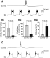
Concentration-dependent actions of octopamine (OA) on B67 burst parameters. Experiments were conducted in a medium (raised concentrations of Ca++ and Mg++) that attenuates chemical synaptic signaling. (A) OA produced multiple effects on B67 burst parameters that were reversible with wash. Bath OA application (1×10⁻⁵ mol l−1) resulted in an increase in the number of impulses per burst, burst duration and intraburst spike frequency of B67. All burst parameters returned to control levels following wash (30 min) with OA-free solution. (B) Concentration-dependence of OA actions on the B67 burst parameters. (Bi) Recordings show representative control, OA 10−7 mol l−1 and OA 10−4 mol l−1 bursts. (Bii) Dose–response curve based on pooled experiments (N=5) in which OA was applied in increasing concentrations over four orders of magnitude. The EC50 for this effect was estimated to be approximately 1×10⁻⁵ mol l−1 (broken lines). (C) Group data demonstrating effects of OA on B67 burst parameters. Comparison of parameters measured prior to OA application with those observed in the presence of the EC50, 1×10⁻⁵ mol l−1. Significant increases were observed in (Ci) the number of bursts per minute (control: 2.3±0.4; OA: 6.5±0.2; P<0.05; N=6), (Cii) the burst duration (control: 0.99±0.07 s; OA: 1.58±0.18 s; P<0.05; N=6) and (Ciii) the spikes per burst (control: 11.4±0.9 impulses s−1; OA: 31.8±3.8 impulses s−1; t=5.27; N=6; P<0.05).
Fig. 5.
Fig. 5.
Effects of octopamine (OA) on the B67 driver potential (DP). Experiments were performed in raised divalent solution and tetrodotoxin (TTX) (1×10⁻⁵ mol l−1). (A) Representative B67 recordings show spontaneous DPs. Note that three to four impulses persisted during the rising phase of the control DP in TTX (see Serrano and Miller, 2006). Bath application of OA (1×10⁻⁵ mol l−1) in the presence of TTX prolonged the DP (middle record). This action was reversed following 30 min wash with OA-free solution (bottom record). (Bi) Group data show that DP durations increased from control values of 0.99±0.08 s to 3.49±0.44 s by 1×10−5 mol l−1 OA (P<0.01; N=4). (Bii) OA produced a small increase the amplitude of spontaneous DPs from control values of 21.0±2.1 mV to 24.6±1.6 mV. This increase was not statistically significant (see text).
Fig. 6.
Fig. 6.
Effects of octopamine (OA) on membrane properties of B67. (A) Application of OA (1×10⁻⁵ mol l−1; right panel) did not produce detectable effects on the interburst membrane potential or input resistance of B67. At this concentration, OA produced significant increases in the burst duration and number of impulses per burst (compare with control burst, left panel; see also Fig. 4) but did not alter the basal interburst membrane potential (Vm) (−50 mV in this experiment). Similarly, measurements of the membrane input resistance, tested with hyperpolarizing pulses (5 nA, 1 s) did not disclose effects of OA. (B) The depolarizing relaxation, or ‘sag’, observed in the response of B67 to larger (10–20 nA) and longer (5 s) pulses were increased by OA (compare OA and control panels). To facilitate comparisons, the Vm was stepped to a fixed level (−90 mV; indicated by broken lines), and the sag was measured as the depolarization from this Vm at the termination of the 5 s pulse. The response of B67 immediately following such pulses was also influenced by OA. The number of spikes following the pulse was increased by OA, and the latency to the first impulse was decreased. (C) Quantification of each of these effects with group data demonstrated that (Ci) sag potentials were significantly increased by 10−5 mol l−1 OA (control: 10.1±0.7 mV; OA: 16.4±2.1 mV; P<0.05; N=5); (Cii) the number of impulses following pulses was increased (control: 7.6±2.6; OA: 15.7±2.5; P<0.05; N=5); and (Ciii) the latency to the initial spike following the pulses was decreased in OA (control: 1.30±0.37 s; OA: 0.42±0.07 s; P<0.05; N=5)
Fig. 7.
Fig. 7.
Octopamine (OA) increases the frequency and regularity of B67 bursting. (A) Under control conditions (control, raised divalent solution), B67 bursting was infrequent and irregular. In addition to its actions on parameters of each burst, OA also increased the frequency and regularity of the B67 bursting. (Bi) Quantification with group data suggested that OA could decrease in the interburst interval (IBI) (mean IBI control: 93.3 s, OA: 11.6 s) but this effect (P=0.23) did not did not satisfy our criterion for statistical significance due to the large dispersion of control values (±45.3 s). (Bii) Normalizing the IBI dispersions by calculating their coefficients of variation revealed a significant reduction by OA (control: 88.4±8.9%; OA: 21.1±4.7%; P<0.001; N=6). (Biii) The decreased IBIs and increased burst durations contributed to increasing the B67 duty cycle in the presence of OA (control: 2.4±0.2%; OA: 11.4±2.3%; P<0.05; N=5). (C) OA-induced rhythmicity persisted in the presence of tetrodotoxin (TTX). In TTX (1×10⁻⁵ mol l−1; high divalent solution; control), spontaneous B67 driver potentials (DPs) occurred in an irregular and infrequent fashion. Application of OA (1×10⁻⁴ mol l−1; OA) caused the DPs to occur rhythmically.
Fig. 8.
Fig. 8.
Octopamine (OA) increases the rhythmicity of bursting in the two B67s but does not increase their synchrony. (Ai) Left panel; simultaneous recordings from the B67 neuron in the left buccal hemiganglion (LB67) and in the right hemiganglion (RB67) under control conditions (raised divalent medium). Right panel: addition of OA (1×10⁻⁴ mol l−1) resulted in both B67s becoming highly rhythmic. (Aii, Aiii) Left panels: autocorrelograms generated with data from the two B67s exhibited very weak rhythmicity within the time window examined (±5 min; bin width: 10 s). Right panels: in OA, the autocorrelograms for both B67s showed very strong rhythmicity with periodicities in the range of 8–10 s. Note that the time window of the OA autocorrelograms was only ±1 min due to the high frequency of burst activity (bin width: 2 s). (Bi) Comparison of IBIs (15 from each B67) from experiment shown in A. Although bursting of both B67s was highly rhythmic in OA, their rates were different, with the left B67 (range of IBIs: 8.5–9.2 s) bursting slightly slower than the right B67 (range of IBIs: 8.0–8.5 s). (Bii) The index of synchrony, calculated as the proportion of B67 bursts that exhibited overlap with contralateral firing (see Materials and methods) was slightly, but not significantly, increased by OA (control: 0.10±0.04; OA: 0.19±0.04; P=0.175; N=4). (Biii) The cross-correlogram of LB67 and RB67 bursting confirmed their lack of synchrony (time window: ±1 min; bin width: 2 s).
Fig. 9.
Fig. 9.
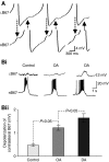
Modulators regulate perturbations of membrane potential between contralateral B67s. (A) Recordings from the left (LB67, upper record) and right (RB67, lower record) shown at high gain [octopamine (OA), 1×10⁻⁴ mol l−1]. In both recordings, bursts were truncated (note discontinuous records) to enable viewing small (approximately 1 mV) depolarizations in contralateral cells (arrows). (B) Comparison of the slow depolarizations produced in B67 during bursts in its contralateral counterpart. (Bi) Augmentation of the depolarization in RB67 produced by OA (1×10⁻⁴ mol l−1) was less than that produced by dopamine (DA; 1×10⁻⁴ mol l−1). (Bii) Group data comparing the effects of OA and DA on the contralateral depolarization produced by B67 bursting. All experiments (N=7; OA applied first in four experiments, DA in three experiments) were conducted in high divalent solution. While the depolarizations measured in the presence of OA (1.22±0.11 mV) were larger than control values (0.47±0.05 mV; t=4.237; N=7; P<0.05), those observed during application of DA were significantly larger than in OA (1.64±0.18 mV; t=2.373; N=7; P<0.05).
Fig. 10.
Fig. 10.
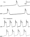
Effects of octopamine (OA) on B67 relative burst timing are not dependent on impulse-mediated signaling. Under control conditions [tetrodotoxin (TTX), 1×10⁻⁵ mol l−1], the left B67 (LB67) and right B67 (RB67) neurons produced spontaneous arrhythmic and asynchronous driver potentials (DPs). Addition of OA (TTX + OA, 1×10⁻⁴ mol l−1) increased the frequency and regularity of spontaneous DPs. OA did not impose synchrony between the DPs of the left and right B67s.
Fig. 11.
Fig. 11.
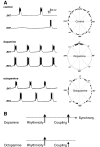
Common and distinct modulatory actions of octopamine (OA) and dopamine (DA) on B67 bursting. (A) Sequential application of DA and OA illustrate their common and distinct actions on B67 burst activity. Left panels: while DA and OA both produce increases in the duration, frequency and rhythmicity of B67 bursting, only DA also imposes synchronous bilateral B67 bursting. Right panels: circular plots illustrate phase relations between the two B67 neurons under control conditions (top), in DA (middle) and in OA (bottom). (B) Schematic interpretation of proposed modulator actions on B67. It is hypothesized that synchrony of bursting between the two B67s requires that their rhythmicity and mutual coupling both exceed a critical level (horizontal broken lines). Although DA and OA both exert comparable increases in the rhythmicity of B67 bursting (compare left vertical arrows), only DA enhances their coupling sufficiently to achieve burst synchrony (compare right vertical arrows).

Similar articles

Cited by

References

    1. Angstadt J. D., Calabrese R. L. (1989). A hyperpolarization-activated inward current in heart interneurons of the medicinal leech. J. Neurosci. 9, 2846-2857 - PMC - PubMed
    1. Arbas E. D., Calabrese R. L. (1987). Ionic conductances underlying the activity of interneurons that control heartbeat in the medicinal leech. J. Neurosci. 7, 3945-3952 - PMC - PubMed
    1. Benson J. A. (1984). Octopamine alters rhythmic activity in the isolated cardiac ganglion of the crab, Portunus sanguinolentus. Neurosci. Lett. 44, 59-64 - PubMed
    1. Berry M. D. (2007). The potential of trace amines and their receptors for treating neurological and psychiatric diseases. Rev. Recent Clin. Trials 2, 3-19 - PubMed
    1. Borovikov D., Evans C. G., Jing J., Rosen S. C., Cropper E. C. (2000). A proprioceptive role for an exteroceptive mechanoafferent neuron in Aplysia. J. Neurosci. 20, 1990-2002 - PMC - PubMed

Publication types