

A new paradigm of bacteria-gut interplay brought through the study of Shigella
- PMID: 20228623
- PMCID: PMC3417848
- DOI: 10.2183/pjab.86.229
A new paradigm of bacteria-gut interplay brought through the study of Shigella
Abstract
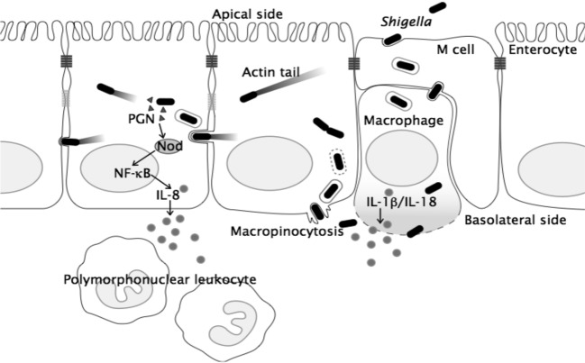
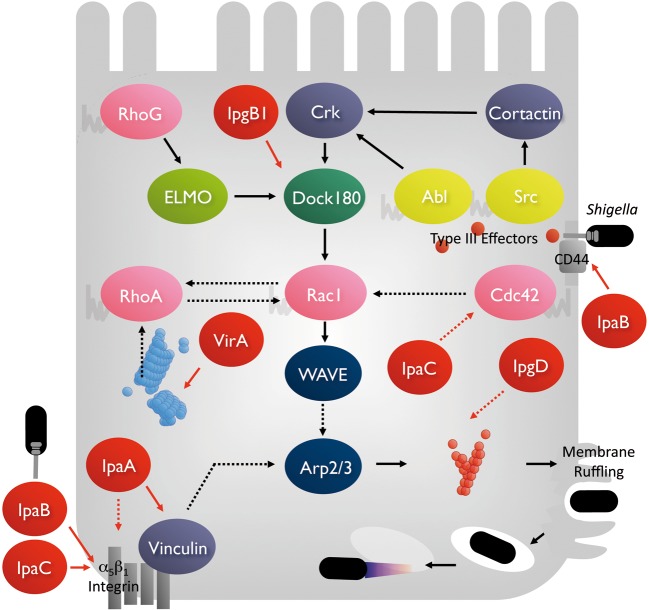
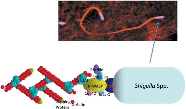
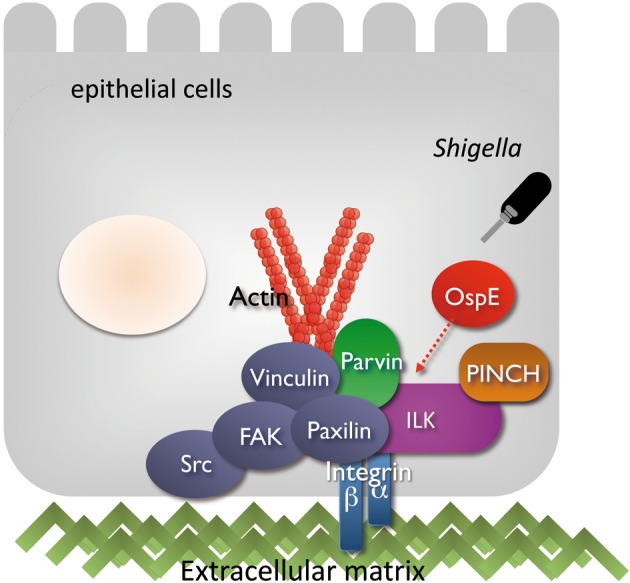
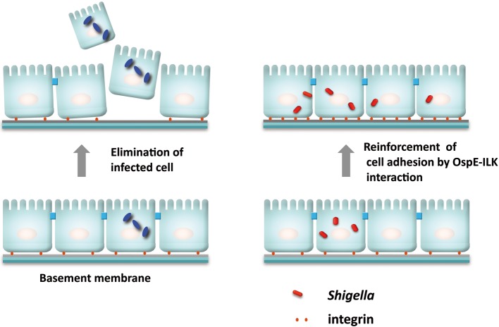
Bacteria-gut epithelial interplay and the mucosal immune response are the most critical issues in determining the fate of bacterial infection and the severity of diseases. Shigella species (abbreviated here as Shigella), the causative agent of bacillary dysentery (shigellosis), are highly adapted human pathogens that are capable of invading and colonizing the intestinal epithelium, which results in severe inflammatory colitis. Shigella secrete a large and diverse number (more then 50) of effectors via the type III secretion system (TTSS) during infection, some of which are delivered into the surrounding bacterial space and some others into the host cell cytoplasm and nucleus. The delivered effectors mimic and usurp the host cellular functions, and modulate host cell signaling and immune response, thus playing pivotal roles in promoting bacterial infection and circumventing host defense systems. This article overviews the pathogenic characteristics of Shigella, and highlights current topics related to the bacterial infectious stratagem executed by the TTSS-secreted effectors. Though bacterial stratagems and the molecular mechanisms of infection vary greatly among pathogens, the current studies of Shigella provide a paradigm shift in bacterial pathogenesis.
Figures

Similar articles
-
[Pathogenesis of Shigella: the study of bacteria-host interplay at the intestinal mucosal barriers].Nihon Saikingaku Zasshi. 2012;67(4):257-68. doi: 10.3412/jsb.67.257. Nihon Saikingaku Zasshi. 2012. PMID: 23269180 Review. Japanese.
-
Shigella infection of intestinal epithelium and circumvention of the host innate defense system.Curr Top Microbiol Immunol. 2009;337:231-55. doi: 10.1007/978-3-642-01846-6_8. Curr Top Microbiol Immunol. 2009. PMID: 19812985 Review.
-
The versatility of Shigella effectors.Nat Rev Microbiol. 2008 Jan;6(1):11-6. doi: 10.1038/nrmicro1814. Nat Rev Microbiol. 2008. PMID: 18059288 Review.
-
Shigella are versatile mucosal pathogens that circumvent the host innate immune system.Curr Opin Immunol. 2011 Aug;23(4):448-55. doi: 10.1016/j.coi.2011.06.001. Epub 2011 Jul 15. Curr Opin Immunol. 2011. PMID: 21763117 Review.
-
Cellular Aspects of Shigella Pathogenesis: Focus on the Manipulation of Host Cell Processes.Front Cell Infect Microbiol. 2016 Mar 31;6:38. doi: 10.3389/fcimb.2016.00038. eCollection 2016. Front Cell Infect Microbiol. 2016. PMID: 27066460 Free PMC article. Review.
Cited by
-
Always one step ahead: How pathogenic bacteria use the type III secretion system to manipulate the intestinal mucosal immune system.J Inflamm (Lond). 2011 May 3;8:11. doi: 10.1186/1476-9255-8-11. J Inflamm (Lond). 2011. PMID: 21539730 Free PMC article.
-
Post-infectious irritable bowel syndrome: mechanistic insights into chronic disturbances following enteric infection.World J Gastroenterol. 2014 Apr 14;20(14):3976-85. doi: 10.3748/wjg.v20.i14.3976. World J Gastroenterol. 2014. PMID: 24744587 Free PMC article. Review.
-
Bacterial nucleomodulins: A coevolutionary adaptation to the eukaryotic command center.PLoS Pathog. 2021 Jan 21;17(1):e1009184. doi: 10.1371/journal.ppat.1009184. eCollection 2021 Jan. PLoS Pathog. 2021. PMID: 33476322 Free PMC article. Review.
-
A Brief History of Shigella.EcoSal Plus. 2018 Jan;8(1):10.1128/ecosalplus.ESP-0006-2017. doi: 10.1128/ecosalplus.ESP-0006-2017. EcoSal Plus. 2018. PMID: 29318984 Free PMC article. Review.
-
Bacterial Actin-Specific Endoproteases Grimelysin and Protealysin as Virulence Factors Contributing to the Invasive Activities of Serratia.Int J Mol Sci. 2020 Jun 4;21(11):4025. doi: 10.3390/ijms21114025. Int J Mol Sci. 2020. PMID: 32512842 Free PMC article. Review.
References
-
- Ogawa M., Handa Y., Ashida H., Suzuki M., Sasakawa C. (2008) The versatility of Shigella effectors. Nat. Rev. Microbiol. 6, 11–16 - PubMed
-
- Ashida H., Ogawa M., Mimuro H., Sasakawa C. (2009) Shigella infection of intestinal epithelium and circumvention of the host innate defense system. Curr. Top. Microbiol. Immunol. 337, 231– 255 - PubMed
-
- Shiga K. (1898) Ueber den errenger der dysenterie in Japan. Zent. Bakteriol. Microbiol. Hyg. 23, 599–600
-
- Trofa A.F., Ueno-Olsen H., Oiwa R., Yoshikawa M. (1999) Dr. Kiyoshi Shiga: discovery of the dysentery bacillus. Clin. Infect. Dis. 29, 1303–1306 - PubMed
-
- DuPont H.L., Levine M.M., Hornick R.B., Formal S.B. (1989) Inoculum size in shigellosis and implications for expected mode of transmission. J. Infect. Dis. 159, 1126–1128 - PubMed