. 2010 Apr;11(4):295-302.
doi: 10.1038/ni.1855. Epub 2010 Mar 14.

An intravascular immune response to Borrelia burgdorferi involves Kupffer cells and iNKT cells

Affiliations

An intravascular immune response to Borrelia burgdorferi involves Kupffer cells and iNKT cells

Woo-Yong Lee et al. Nat Immunol. 2010 Apr.

Abstract

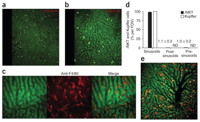
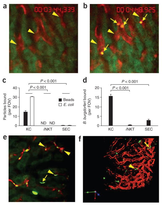
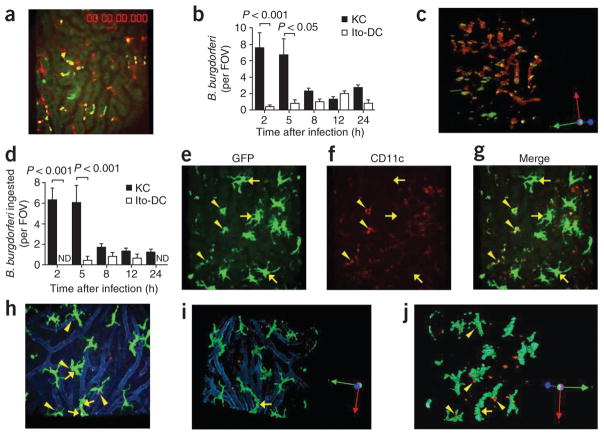
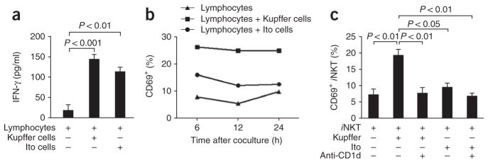
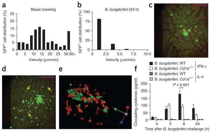
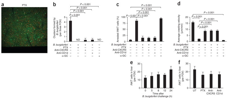
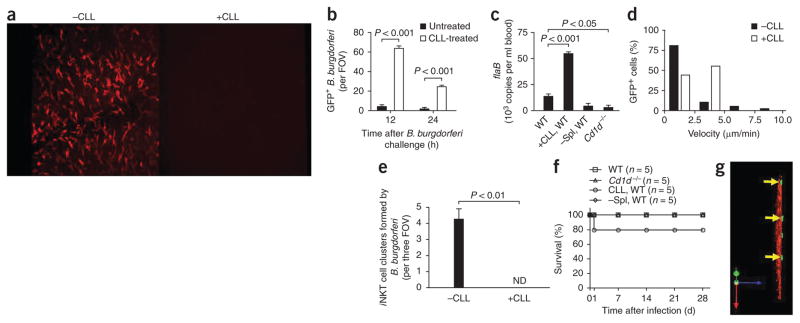
Here we investigate the dynamics of the hepatic intravascular immune response to a pathogen relevant to invariant natural killer T cells (iNKT cells). Immobilized Kupffer cells with highly ramified extended processes into multiple sinusoids could effectively capture blood-borne, disseminating Borrelia burgdorferi, creating a highly efficient surveillance and filtering system. After ingesting B. burgdorferi, Kupffer cells induced chemokine receptor CXCR3-dependent clustering of iNKT cells. Kupffer cells and iNKT cells formed stable contacts via the antigen-presenting molecule CD1d, which led to iNKT cell activation. An absence of iNKT cells caused B. burgdorferi to leave the blood and enter the joints more effectively. B. burgdorferi that escaped Kupffer cells entered the liver parenchyma and survived despite Ito cell responses. Kupffer cell-iNKT cell interactions induced a key intravascular immune response that diminished the dissemination of B. burgdorferi.


Conflict of interest statement

The authors declare no competing financial interests.

Figures

Figure 1
Figure 1
Distribution of iNKT cells and Kupffer cells in the hepatic microvasculature. Spinning-disk confocal intravital microscopy of the vasculature of Cxcr6gfp/+ and BALB/c mouse livers. (a,b) CXCR6+ cells in the liver of Cxcr6gfp/+ mice. (c) Liver-specific Kupffer cells (red) labeled with phycoerythrin-conjugated anti-F4/80 in the hepatic sinusoids of a BALB/c mouse. (d) GFP+ cells and F4/80+ cells in the sinusoids, post-sinusoidal venules and pre-sinusoidal venules of Cxcr6gfp/+ mouse livers (n = 7 mice) in 21 FOV (iNKT cells) or 10 FOV (Kupffer cells). Numbers in graph indicate percent iNKT cells per FOV for bars not visible; ND, not detected. (e) Dextran conjugated to tetramethylrhodamine (2 megadalton; red) and polychromatic microspheres (green and yellow) bound to Kupffer cells. Original magnification, ×4 (a and iNKT cells in d), ×10 (b,e and Kupffer cells in d) or ×20 (c). Data are representative of more than three independent experiments (ac,e) or seven experiments (d; error bars, s.e.m.).
Figure 2
Figure 2
Binding capacity of Kupffer cells, iNKT cells and SECs for beads and bacteria. (a,b) Microscopy of red Kupffer cells (arrowheads) before (a) and after (b) intravenous injection of polychromatic microspheres (arrows, b) into BALB/c mice. (c,d) Quantification of the binding of beads or E. coli (c) or B. burgdorferi (d) to Kupffer cells (KC), iNKT cells or SECs in Cxcr6gfp/+ mice given intravenous injection of polychromatic microspheres, or E. coli (c) or B. burgdorferi (d) expressing GFP or labeled with the red fluorescent nucleic acid stain Syto 60. (e) Binding of B. burgdorferi to the endothelium (arrow) as well as to Kupffer cells (arrowheads) in the hepatic sinusoids of a mouse treated as described in d (differences in activity, Supplementary Video 5). (f) Microcopy of B. burgdorferi in the liver of a mouse treated as described in d; yellow arrowhead indicates a GFP+ spirochete in the process of emigrating, and yellow arrow indicates a spirochete that has left the vasculature. Original magnification, ×20 (a,b,e,f). P values (c,d), Bonferroni’s multiple-comparison test. Data are representative of more than three independent experiments (ae) or two experiments (f).
Figure 3
Figure 3
Ingestion of B. burgdorferi by Kupffer cells and Ito cells. (a) Visualization of the hepatic vasculature of a Cxcr6gfp/+ mouse at 2 h after the administration of GFP-expressing B. burgdorferi (yellow dots, shown interacting with red-labeled Kupffer cells). (b) Association of GFP-expressing or Tomato-expressing B. burgdorferi with Kupffer cells and Ito or dendritic cells (Ito-DC), measured in intravital videos of Cxcr6gfp/+ and Cx3cr1gfp/+ mice, respectively. (c) A z-stack reconstruction of two-dimensional microscopy, showing attachment and ingestion of spirochetes by Kupffer cells (rotation, Supplementary Fig. 1). (d) Total ingested B. burgdorferi confirmed after 360° rotation of z-stack-reconstructed images. (eg) CD11c, GFP+ big stellate (Ito) cells (arrows) and relatively small CD11c+GFP+ (dendritic) cells (arrowheads) in Cx3cr1gfp/+ mouse liver. (h) Tomato-expressing B. burgdorferi bound by GFP+ Ito cells in Cx3cr1gfp/+ mouse liver. Arrows indicate associated spirochetes; arrowheads indicate spirochetes out of Ito cells. Vessels were stained with anti-PECAM-1 (blue). (i,j) Ingestion of Tomato-expressing B. burgdorferi by Ito cells, assessed after z-stack reconstruction. Original magnification, ×20 (a,c,ej). P values (b,d), Bonferroni’s multiple-comparison test. Data are representative of two independent experiments per group with more than four FOV each (ad,hj; error bars (b,d), s.e.m.) or two experiments (eg).
Figure 4
Figure 4
Antigen presentation by Kupffer cells and Ito cells. (a) Release of IFN-γ from liver-derived mixed-lymphocyte populations stimulated for 4 d with Kupffer or Ito cells isolated from BALB/c mice infected by injection of B. burgdorferi. (b,c) Expression of CD69 in mixed-lymphocyte populations treated as described in a (b) and a pure iNKT cell population isolated from Cxcr6gfp/+ mice (Supplementary Methods) and cultured alone or together with Kupffer cells or Ito cells (as described in a) with or without anti-CD1d (c), evaluated by flow cytometry. P values (a,c), Bonferroni’s multiple-comparison test. Data are representative of one experiment (mean and s.e.m. of three to four wells (a) or pooled cells from three to four wells (b)) or three independent experiments (c).
Figure 5
Figure 5
Changes in iNKT cell activity after B. burgdorferi infection. (a,b) GFP+ cell tracks in vehicle-treated mice (a) and at 24 h after injection of B. burgdorferi into Cxcr6gfp/+ mice (b). (c,d) Distribution of GFP+ iNKT cells and red-labeled Kupffer cells at 8 h (c) and 24 h (d) after injection of GFP-expressing B. burgdorferi. Arrows indicate surviving spirochetes. (e) A z-stack reconstruction showing Kupffer cells (red) and iNKT cells (green) at 24 h after infection. Original magnification, ×20 (c,e) or ×10 (d). (f) Enzyme-linked immunosorbent assay of IFN-γ and IL-4 in serum samples obtained from wild-type (WT) and Cd1d−/− mice before or 5, 8 or 24 h after B. burgdorferi injection. P values, Bonferroni’s multiple-comparison test. Data are representative of three independent experiments with more than ten FOV (a) or more than two independent experiments (be) or one experiment with four to six mice per group (f; error bars, s.e.m.).
Figure 6
Figure 6
Inhibition of iNKT cell cluster formation, average crawling velocity and stationary adhesion by pertussis toxin, anti-CXCR3 and anti-CD1d. Analysis of the hepatic microvasculature (visualized by spinning-disk confocal intravital microscopy) and formation of iNKT cell clusters (counted by intravital video) in Cxcr6gfp/+ mice pretreated with pertussis toxin (PTX), anti-CXCR3, anti-CD1d and/or α-GalCer (α-GC) before injection of B. burgdorferi. (a) Distribution of iNKT cells after PTX treatment, assessed 24 h after infection. (b) Cluster formation by B. burgdorferi at 24 h after infection. Original magnification, ×4 (a,b). (c,d) Effect of pretreatment on the average crawling velocity (c) and stationary arrest (d) of iNKT cells at 24 h after infection. (e) Liver iNKT cells during the first 24 h of B. burgdorferi infection. (f) Effect of pretreatment on the number of iNKT cells in the liver sinusoids (n = 4–6 mice per group). Original magnification, ×10 (e,f). P values (bd), Bonferroni’s multiple-comparison test. Data are representative of three experiments (a) or more than two independent experiments per group (bf; error bars, s.e.m.).
Figure 7
Figure 7
Role of Kupffer cells in B. burgdorferi infection. (a) Kupffer cells labeled with anti-F4/80 before (-CLL) and 24 h after (+CLL) intravenous injection of CLLs (administered 24 h before B. burgdorferi infection to deplete mice of Kupffer cells). Original magnification, ×10. (b) Spirochetes remaining in the liver 12 h and 24 h after B. burgdorferi injection in untreated mice (-CLL) and CLL-treated mice (+CLL). (c) Spirochetes remaining in the blood 3 d after B. burgdorferi injection in untreated, CLL-treated and splenectomized (–Spl) wild-type mice and in Cd1d−/− mice, assessed by quantitative PCR analysis of B. burgdorferi flaB. (d) Distribution of iNKT cell velocities at 5 h after spirochete challenge; nonfluorescent B. burgdorferi were administered to avoid possible confusion of GFP-expressing B. burgdorferi with GFP+ iNKT cells in CLL-treated mice. (e,f) Formation of iNKT cell clusters at 24 h after B. burgdorferi infection. (e) Mortality of untreated, CLL-treated and splenectomized (-Spl) wild-type mice and of Cd1d−/− mice after B. burgdorferi infection. (g) Migration of GFP-expressing spirochetes (arrows) out of microvasculature (space of Dissé) in a CLL-treated mouse. Original magnification, ×20. P values Bonferroni’s multiple-comparison test (b) or Student’s t-test (c,e). Data are representative of three experiments (a), more than two independent experiments per group (b,d,e,g), one experiment with duplicate pooled results of three mice (c) or one experiment with five mice per group (f; error bars (b,c,e), s.e.m.).

References

    1. Geissmann F, et al. Intravascular immune surveillance by CXCR6+ NKT cells patrolling liver sinusoids. PLoS Biol. 2005;3:e113. - PMC - PubMed
    1. Auffray C, et al. Monitoring of blood vessels and tissues by a population of monocytes with patrolling behavior. Science. 2007;317:666–670. - PubMed
    1. Kronenberg M. Toward an understanding of NKT cell biology: progress and paradoxes. Annu Rev Immunol. 2005;23:877–900. - PubMed
    1. Kawano T, et al. CD1d-restricted and TCR-mediated activation of Vα14 NKT cells by glycosylceramides. Science. 1997;278:1626–1629. - PubMed
    1. Brossay L, et al. CD1d-mediated recognition of an α-galactosylceramide by natural killer T cells is highly conserved through mammalian evolution. J Exp Med. 1998;188:1521–1528. - PMC - PubMed

Publication types