. 2010 Apr;17(4):398-402.
doi: 10.1038/nsmb.1782. Epub 2010 Mar 14.

An extracellular steric seeding mechanism for Eph-ephrin signaling platform assembly

Affiliations

An extracellular steric seeding mechanism for Eph-ephrin signaling platform assembly

Elena Seiradake et al. Nat Struct Mol Biol. 2010 Apr.

Abstract

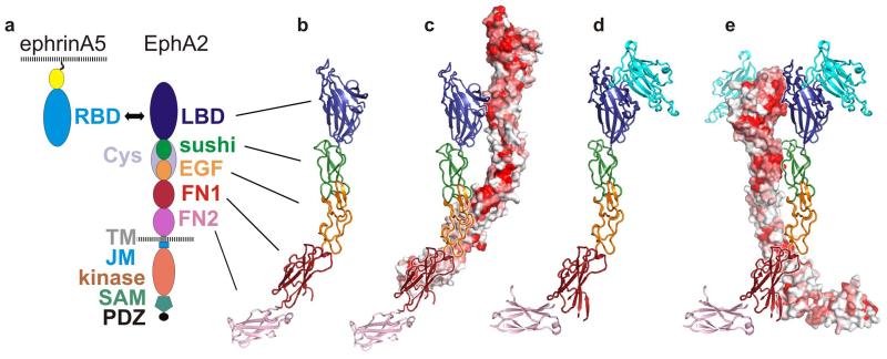
Erythropoetin-producing hepatoma (Eph) receptors are cell-surface protein tyrosine kinases mediating cell-cell communication. Upon activation, they form signaling clusters. We report crystal structures of the full ectodomain of human EphA2 (eEphA2) both alone and in complex with the receptor-binding domain of the ligand ephrinA5 (ephrinA5 RBD). Unliganded eEphA2 forms linear arrays of staggered parallel receptors involving two patches of residues conserved across A-class Ephs. eEphA2-ephrinA5 RBD forms a more elaborate assembly, whose interfaces include the same conserved regions on eEphA2, but rearranged to accommodate ephrinA5 RBD. Cell-surface expression of mutant EphA2s showed that these interfaces are critical for localization at cell-cell contacts and activation-dependent degradation. Our results suggest a 'nucleation' mechanism whereby a limited number of ligand-receptor interactions 'seed' an arrangement of receptors which can propagate into extended signaling arrays.


Figures

Figure 1
Figure 1. Crystal structures of the complete EphA2 extracellular region (eEphA2) alone and in complex with ephrinA5RBD
a, Domain composition of ephrinAs and Eph receptors. b, Crystal structure of eEphA2 comprising EphA2 LBD, Cys, FN1 and FN2 domains. Colours are as in a. The previously uncharacterized Cys domain is composed of a sushi domain (CCP) and an EGF domain. c, Arrangement of two eEphA2 molecules within the crystal. Their contacts bury 2661 Å2 of molecular surface. The front molecule is displayed and oriented as in a. The back molecule is shown in surface view and coloured according to human Eph class A receptor sequence conservation (red = strongest conservation, white = weakest conservation). d, eEphA2 (colours as in a) in complex with ephrinA5RBD (cyan). The orientation of the sushi domain is the same as in b and c. e, Arrangement of neighbouring complexes within the crystal. The eEphA2 molecule on the right is oriented and coloured as in d. Its neighbour on the left is in surface view and coloured according to EphA2 sequence conservation. Their contacts bury 1240 Å2.
Figure 2
Figure 2. The eEphA2-ephrinA5RBD complex: structural changes within eEphA2 and formation of array-like clusters
a, b, Crystal packing of eEphA2-ephrinA5RBD. The ribbon diagrams are coloured as in Fig. 1. The cartoon above each panel illustrates the crystal contacts designated interfaces A-F (dotted lines) for eEphA2 (purple) and ephrinA5RBD (cyan). Panel a shows six symmetry-related eEphA2-ephrinA5RBD complexes forming a parallel array; view is approximately along unit cell axis c. The protein arrangement reflects EphA2 and ephrinA5 interaction in trans, i.e. where the proteins are located on opposing cells. Panel b shows six symmetry-related eEphA2-ephrinA5RBD complexes forming an anti-parallel array, view as in f, but tilted by ~45° around unit cell axis a. The arrangement shows EphA2 and ephrinA5 interacting both in trans and in cis, consistent with both proteins presented on both cells. c-f, Interfaces A-F are shown as black lines. Colours are as in a. Interfaces A-E form the crystal contact interactions shown in a, interface F is a major interface but contributes only to the contacts shown in b.
Figure 3
Figure 3. EphA2 localization at cell-cell contacts depends on ectodomain clustering
Representative results are presented of functional studies using HEK293T cells. A full set of results is provided in Supplementary Fig. 6. Filled white arrows indicate cell-cell contacts between transfected cells. Open arrows point to cell-cell contacts between a transfected cell and a non-transfected cell. a-c, mVenus-tagged EphA2 accumulates at cell-cell contacts of transfected cells. d-e, Mutants in interfaces A, B, D and, to some extent, interface F do not cluster at cell-cell contacts of transfected cells but are distributed evenly on the cell surface (the interface B mutant G131Y is shown as an example). a, d Fluorescence images are shown. b, e, Phase contrast images of the corresponding cells. c, f, Overlay images of a, b, d, e.
Fig.4
Fig.4. A mechanism for the nucleation and propagation of EphA2 signalling assemblies
a, b, Eph and ephrin are shown schematically with ectodomains colour coded as in Fig. 1a and the Eph intracellular region in grey. Lightening flashes indicate the strength of activation. In panel a transient arrays of staggered Ephs snap into in-register signalling arrays on nucleation by ephrins. The intermediate phenotype of interface F mutations in functional assays may reflect weaker interactions between Eph receptors and their ligands, resulting in a weakened signalling activity. In panel b additional cis (interface F) interactions inter-connect anti-parallel arrays further stabilising the signalling assemblies.

References

    1. Pasquale EB. Eph-ephrin bidirectional signaling in physiology and disease. Cell. 2008;133:38–52. - PubMed
    1. Eph Nomenclature Committee Unified nomenclature for Eph family receptors and their ligands, the ephrins. Cell. 1997;90:403–4. - PubMed
    1. Himanen JP, Nikolov DB. Eph signaling: a structural view. Trends Neurosci. 2003;26:46–51. - PubMed
    1. Himanen JP, Saha N, Nikolov DB. Cell-cell signaling via Eph receptors and ephrins. Curr Opin Cell Biol. 2007;19:534–42. - PMC - PubMed
    1. Gale NW, et al. Eph receptors and ligands comprise two major specificity subclasses and are reciprocally compartmentalized during embryogenesis. Neuron. 1996;17:9–19. - PubMed

Publication types