Imaging type-III secretion reveals dynamics and spatial segregation of Salmonella effectors
- PMID: 20228815
- PMCID: PMC2862489
- DOI: 10.1038/nmeth.1437
Imaging type-III secretion reveals dynamics and spatial segregation of Salmonella effectors
Abstract
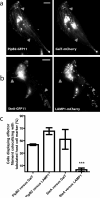
The type-III secretion system (T3SS) enables gram-negative bacteria to inject effector proteins into eukaryotic host cells. Upon entry, T3SS effectors work cooperatively to reprogram host cells, enabling bacterial survival. Progress in understanding when and where effectors localize in host cells has been hindered by a dearth of tools to study these proteins in the native cellular environment. We report a method to label and track T3SS effectors during infection using a split-GFP system. We demonstrate this technique by labeling three effectors from Salmonella enterica (PipB2, SteA and SteC) and characterizing their localization in host cells. PipB2 displayed highly dynamic behavior on tubules emanating from the Salmonella-containing vacuole labeled with both endo- and exocytic markers. SteA was preferentially enriched on tubules localizing with Golgi markers. This segregation suggests that effector targeting and localization may have a functional role during infection.
Figures


Comment in
-
The 'when and whereabouts' of injected pathogen effectors.Nat Methods. 2010 Apr;7(4):267-9. doi: 10.1038/nmeth0410-267. Nat Methods. 2010. PMID: 20354515
Similar articles
-
Salmonella-containing vacuoles display centrifugal movement associated with cell-to-cell transfer in epithelial cells.Infect Immun. 2009 Mar;77(3):996-1007. doi: 10.1128/IAI.01275-08. Epub 2008 Dec 22. Infect Immun. 2009. PMID: 19103768 Free PMC article.
-
Salmonella Effector SteA Suppresses Proinflammatory Responses of the Host by Interfering With IκB Degradation.Front Immunol. 2019 Dec 10;10:2822. doi: 10.3389/fimmu.2019.02822. eCollection 2019. Front Immunol. 2019. PMID: 31921113 Free PMC article.
-
Recent progress in molecular mechanisms of Salmonella effectors involved in gut epithelium invasion.Mol Biol Rep. 2025 Jun 16;52(1):601. doi: 10.1007/s11033-025-10715-9. Mol Biol Rep. 2025. PMID: 40522423 Review.
-
The Salmonella effector SteA binds phosphatidylinositol 4-phosphate for subcellular targeting within host cells.Cell Microbiol. 2016 Jul;18(7):949-69. doi: 10.1111/cmi.12558. Epub 2016 Mar 11. Cell Microbiol. 2016. PMID: 26676327
-
Functions of the Salmonella pathogenicity island 2 (SPI-2) type III secretion system effectors.Microbiology (Reading). 2012 May;158(Pt 5):1147-1161. doi: 10.1099/mic.0.058115-0. Epub 2012 Mar 15. Microbiology (Reading). 2012. PMID: 22422755 Review.
Cited by
-
An Evolutionarily Conserved PLC-PKD-TFEB Pathway for Host Defense.Cell Rep. 2016 May 24;15(8):1728-42. doi: 10.1016/j.celrep.2016.04.052. Epub 2016 May 12. Cell Rep. 2016. PMID: 27184844 Free PMC article.
-
Intracellular Salmonella delivery of an exogenous immunization antigen refocuses CD8 T cells against cancer cells, eliminates pancreatic tumors and forms antitumor immunity.Front Immunol. 2023 Oct 5;14:1228532. doi: 10.3389/fimmu.2023.1228532. eCollection 2023. Front Immunol. 2023. PMID: 37868996 Free PMC article.
-
The motility regulator flhDC drives intracellular accumulation and tumor colonization of Salmonella.J Immunother Cancer. 2019 Feb 12;7(1):44. doi: 10.1186/s40425-018-0490-z. J Immunother Cancer. 2019. PMID: 30755273 Free PMC article.
-
The SdiA-regulated gene srgE encodes a type III secreted effector.J Bacteriol. 2014 Jun;196(12):2301-12. doi: 10.1128/JB.01602-14. Epub 2014 Apr 11. J Bacteriol. 2014. PMID: 24727228 Free PMC article.
-
What the SIF Is Happening-The Role of Intracellular Salmonella-Induced Filaments.Front Cell Infect Microbiol. 2017 Jul 25;7:335. doi: 10.3389/fcimb.2017.00335. eCollection 2017. Front Cell Infect Microbiol. 2017. PMID: 28791257 Free PMC article. Review.
References
-
- Johnson S, Deane JE, Lea SM. The type III needle and the damage done. Curr.Opin.Struct.Biol. 2005;15:700–707. - PubMed
-
- Boucrot E, Beuzon CR, Holden DW, Gorvel JP, Meresse S. Salmonella typhimurium SifA effector protein requires its membrane-anchoring C-terminal hexapeptide for its biological function. J.Biol.Chem. 2003;278:14196–14202. - PubMed
Publication types
MeSH terms
Substances
Grants and funding
LinkOut - more resources
Full Text Sources
Other Literature Sources
Medical
Research Materials

