Src family kinase/abl inhibitor dasatinib suppresses proliferation and enhances differentiation of osteoblasts
- PMID: 20228840
- PMCID: PMC2921959
- DOI: 10.1038/onc.2010.73
Src family kinase/abl inhibitor dasatinib suppresses proliferation and enhances differentiation of osteoblasts
Abstract
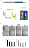
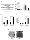
Dasatinib, a dual Src family kinase and Abl inhibitor, is being tested clinically for the treatment of prostate cancer bone metastasis. Bidirectional interactions between osteoblasts and prostate cancer cells are critical in the progression of prostate cancer in bone, but the effect of dasatinib on osteoblasts is unknown. We found that dasatinib inhibited proliferation of primary mouse osteoblasts isolated from mouse calvaria and the immortalized MC3T3-E1 cell line. In calvarial osteoblasts from Col-luc transgenic mice carrying osteoblast-specific Col1alpha1 promoter reporter, luciferase activity was inhibited. Dasatinib also inhibited fibroblast growth factor-2-induced osteoblast proliferation, but strongly promoted osteoblast differentiation, as reflected by stimulation of alkaline phosphatase activity, osteocalcin secretion and osteoblast mineralization. To determine how dasatinib blocks proliferative signaling in osteoblasts, we analyzed the expression of a panel of tyrosine kinases, including Src, Lyn, Fyn, Yes and Abl, in osteoblasts. In the Src family kinases, only Src was activated at a high level. Abl was expressed at a low level in osteoblasts. Phosphorylation of Src-Y419 or Abl-Y245 was inhibited by dasatinib treatment. Knockdown of either Src or Abl by lenti-shRNA in osteoblasts enhances osteoblast differentiation, suggesting that dasatinib enhances osteoblast differentiation through inhibition of both Src and Abl.
Conflict of interest statement
There is no conflict of interest from all authors.
Figures


Similar articles
-
The Src inhibitor dasatinib accelerates the differentiation of human bone marrow-derived mesenchymal stromal cells into osteoblasts.BMC Cancer. 2010 Jun 17;10:298. doi: 10.1186/1471-2407-10-298. BMC Cancer. 2010. PMID: 20565769 Free PMC article.
-
Inhibition of cell growth and up-regulation of MAD2 in human oesophageal squamous cell carcinoma after treatment with the Src/Abl inhibitor dasatinib.Clin Sci (Lond). 2012 Jan;122(1):13-24. doi: 10.1042/CS20110230. Clin Sci (Lond). 2012. PMID: 21751967
-
Dasatinib inhibits site-specific tyrosine phosphorylation of androgen receptor by Ack1 and Src kinases.Oncogene. 2010 Jun 3;29(22):3208-16. doi: 10.1038/onc.2010.103. Epub 2010 Apr 12. Oncogene. 2010. PMID: 20383201 Free PMC article.
-
Inhibition of SRC family kinases and receptor tyrosine kinases by dasatinib: possible combinations in solid tumors.Clin Cancer Res. 2011 Sep 1;17(17):5546-52. doi: 10.1158/1078-0432.CCR-10-2616. Epub 2011 Jun 13. Clin Cancer Res. 2011. PMID: 21670084 Review.
-
Targeting ABL and SRC kinases in chronic myeloid leukemia: experience with dasatinib.Future Oncol. 2006 Dec;2(6):655-65. doi: 10.2217/14796694.2.6.655. Future Oncol. 2006. PMID: 17155893 Review.
Cited by
-
The tyrosine kinase inhibitor dasatinib (SPRYCEL) inhibits chondrocyte activity and proliferation.Blood Cancer J. 2011 Feb;1(2):e2. doi: 10.1038/bcj.2011.1. Epub 2011 Feb 4. Blood Cancer J. 2011. PMID: 22829108 Free PMC article. No abstract available.
-
The Src inhibitor dasatinib accelerates the differentiation of human bone marrow-derived mesenchymal stromal cells into osteoblasts.BMC Cancer. 2010 Jun 17;10:298. doi: 10.1186/1471-2407-10-298. BMC Cancer. 2010. PMID: 20565769 Free PMC article.
-
Autophagy as a modulator and target in prostate cancer.Nat Rev Urol. 2014 Sep;11(9):508-16. doi: 10.1038/nrurol.2014.196. Epub 2014 Aug 19. Nat Rev Urol. 2014. PMID: 25134829 Free PMC article. Review.
-
The Src family kinase inhibitor dasatinib delays pain-related behaviour and conserves bone in a rat model of cancer-induced bone pain.Sci Rep. 2017 Jul 6;7(1):4792. doi: 10.1038/s41598-017-05029-1. Sci Rep. 2017. PMID: 28684771 Free PMC article.
-
Exosomes from Placenta-Derived Mesenchymal Stem Cells Are Involved in Liver Regeneration in Hepatic Failure Induced by Bile Duct Ligation.Stem Cells Int. 2020 Oct 9;2020:5485738. doi: 10.1155/2020/5485738. eCollection 2020. Stem Cells Int. 2020. PMID: 33133194 Free PMC article.
References
-
- Araujo J, Logothetis C. Targeting Src signaling in metastatic bone disease. Int J Cancer. 2009;124:1–6. - PubMed
-
- Aubin JE. Advances in the osteoblast lineage. Biochem Cell Biol. 1998;76:899–910. - PubMed
-
- Bellows CG, Aubin JE. Determination of numbers of osteoprogenitors present in isolated fetal rat calvaria cells in vitro. Dev Biol. 1989;133:8–13. - PubMed
-
- Berman E, Nicolaides M, Maki RG, Fleisher M, Chanel S, Scheu K, et al. Altered bone and mineral metabolism in patients receiving imatinib mesylate. N Engl J Med. 2006;354:2006–2013. - PubMed
Publication types
MeSH terms
Substances
Grants and funding
LinkOut - more resources
Full Text Sources
Molecular Biology Databases
Miscellaneous

