

Metastatic breast tumors express increased tau, which promotes microtentacle formation and the reattachment of detached breast tumor cells
- PMID: 20228842
- PMCID: PMC3132415
- DOI: 10.1038/onc.2010.68
Metastatic breast tumors express increased tau, which promotes microtentacle formation and the reattachment of detached breast tumor cells
Abstract
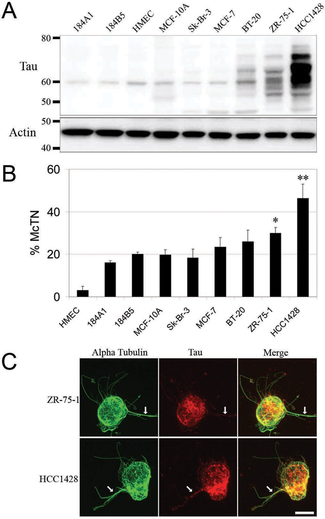
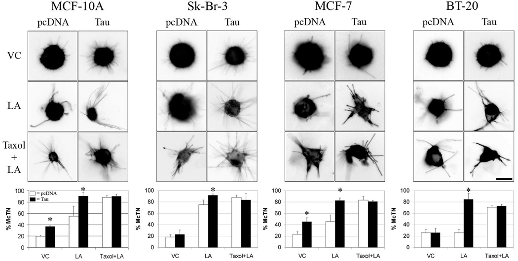
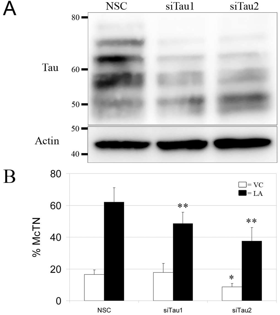
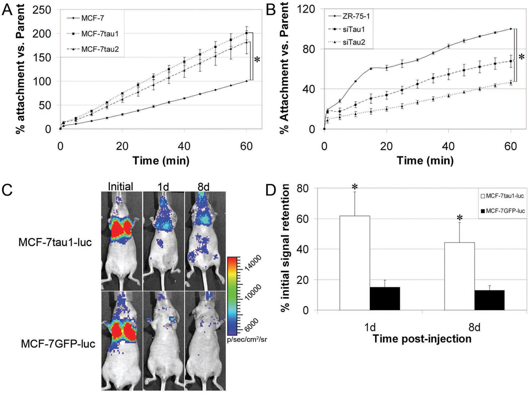
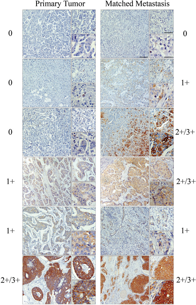
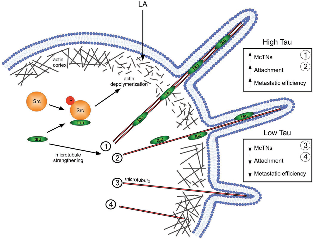
The cytoskeletal organization of detached and circulating tumor cells (CTCs) is currently not well defined and may provide potential targets for new therapies to limit metastatic tumor spread. In vivo, CTCs reattach in distant tissues by a mechanism that is tubulin-dependent and suppressed by polymerized actin. The cytoskeletal mechanisms that promote reattachment of CTCs match exactly with the mechanisms supporting tubulin microtentacles (McTN), which we have recently identified in detached breast tumor cells. In this study, we aimed to investigate how McTN formation is affected by the microtubule-associated protein, tau, which is expressed in a subset of chemotherapy-resistant breast cancers. We demonstrate that endogenous tau protein localizes to McTNs and is both necessary and sufficient to promote McTN extension in detached breast tumor cells. Tau-induced McTNs increase reattachment of suspended cells and retention of CTCs in lung capillaries. Analysis of patient-matched primary and metastatic tumors reveals that 52% possess tau expression in metastases and 26% display significantly increased tau expression over disease progression. Tau enrichment in metastatic tumors and the ability of tau to promote tumor cell reattachment through McTN formation support a model in which tau-induced microtubule stabilization provides a selective advantage during tumor metastasis.
Conflict of interest statement
Figures
Similar articles
-
Curcumin targets breast cancer stem-like cells with microtentacles that persist in mammospheres and promote reattachment.Cancer Res. 2014 Feb 15;74(4):1250-60. doi: 10.1158/0008-5472.CAN-13-1778. Epub 2013 Dec 26. Cancer Res. 2014. PMID: 24371229 Free PMC article.
-
Vimentin filaments support extension of tubulin-based microtentacles in detached breast tumor cells.Cancer Res. 2008 Jul 15;68(14):5678-88. doi: 10.1158/0008-5472.CAN-07-6589. Cancer Res. 2008. PMID: 18632620 Free PMC article.
-
Local anesthetics inhibit kinesin motility and microtentacle protrusions in human epithelial and breast tumor cells.Breast Cancer Res Treat. 2011 Oct;129(3):691-701. doi: 10.1007/s10549-010-1239-7. Epub 2010 Nov 11. Breast Cancer Res Treat. 2011. PMID: 21069453 Free PMC article.
-
Microtentacles tip the balance of cytoskeletal forces in circulating tumor cells.Cancer Res. 2010 Oct 15;70(20):7737-41. doi: 10.1158/0008-5472.CAN-10-1569. Epub 2010 Oct 5. Cancer Res. 2010. PMID: 20924109 Free PMC article. Review.
-
Tubulin Complexity in Cancer and Metastasis.Adv Exp Med Biol. 2024;1452:21-35. doi: 10.1007/978-3-031-58311-7_2. Adv Exp Med Biol. 2024. PMID: 38805123 Review.
Cited by
-
Microtubule disruption reduces metastasis more effectively than primary tumor growth.Breast Cancer Res. 2022 Feb 14;24(1):13. doi: 10.1186/s13058-022-01506-2. Breast Cancer Res. 2022. PMID: 35164808 Free PMC article.
-
Alzheimer's disease and related tauopathies: disorders of disrupted neuronal identity.Trends Neurosci. 2023 Oct;46(10):797-813. doi: 10.1016/j.tins.2023.07.006. Epub 2023 Aug 15. Trends Neurosci. 2023. PMID: 37591720 Free PMC article. Review.
-
Molecular Pathways: New Signaling Considerations When Targeting Cytoskeletal Balance to Reduce Tumor Growth.Clin Cancer Res. 2015 Dec 1;21(23):5209-5214. doi: 10.1158/1078-0432.CCR-15-0328. Epub 2015 Oct 13. Clin Cancer Res. 2015. PMID: 26463706 Free PMC article. Review.
-
Microtubule Hyperacetylation Enhances KL1-Dependent Micronucleation under a Tau Deficiency in Mammary Epithelial Cells.Int J Mol Sci. 2018 Aug 23;19(9):2488. doi: 10.3390/ijms19092488. Int J Mol Sci. 2018. PMID: 30142893 Free PMC article.
-
Blocking the attachment of cancer cells in vivo with DNA aptamers displaying anti-adhesive properties against the carcinoembryonic antigen.Mol Oncol. 2013 Aug;7(4):799-811. doi: 10.1016/j.molonc.2013.03.005. Epub 2013 Apr 11. Mol Oncol. 2013. PMID: 23656757 Free PMC article.
References
-
- Atienza JM, Yu N, Kirstein SL, Xi B, Wang X, Xu X, et al. Dynamic and label-free cell-based assays using the real-time cell electronic sensing system. Assay Drug Dev Technol. 2006;4:597–607. - PubMed
-
- Bhat KM, Setaluri V. Microtubule-associated proteins as targets in cancer chemotherapy. Clin Cancer Res. 2007;13:2849–2854. - PubMed
-
- Bulic B, Pickhardt M, Schmidt B, Mandelkow EM, Waldmann H, Mandelkow E. Development of tau aggregation inhibitors for Alzheimer's disease. Angew Chem Int Ed Engl. 2009;48:1740–1752. - PubMed
-
- Camara O, Rengsberger M, Egbe A, Koch A, Gajda M, Hammer U, et al. The relevance of circulating epithelial tumor cells (CETC) for therapy monitoring during neoadjuvant (primary systemic) chemotherapy in breast cancer. Ann Oncol. 2007;18:1484–1492. - PubMed
Publication types
MeSH terms
Substances
Grants and funding
LinkOut - more resources
Full Text Sources
Other Literature Sources
Medical
