. 2009 Aug;48(8):8JA03-8JA0310.
doi: 10.1143/JJAP.48.08JA03.

Investigation of SNARE-Mediated Membrane Fusion Mechanism Using Atomic Force Microscopy

Affiliations

Investigation of SNARE-Mediated Membrane Fusion Mechanism Using Atomic Force Microscopy

Midhat H Abdulreda et al. Jpn J Appl Phys (2008). 2009 Aug.

Abstract

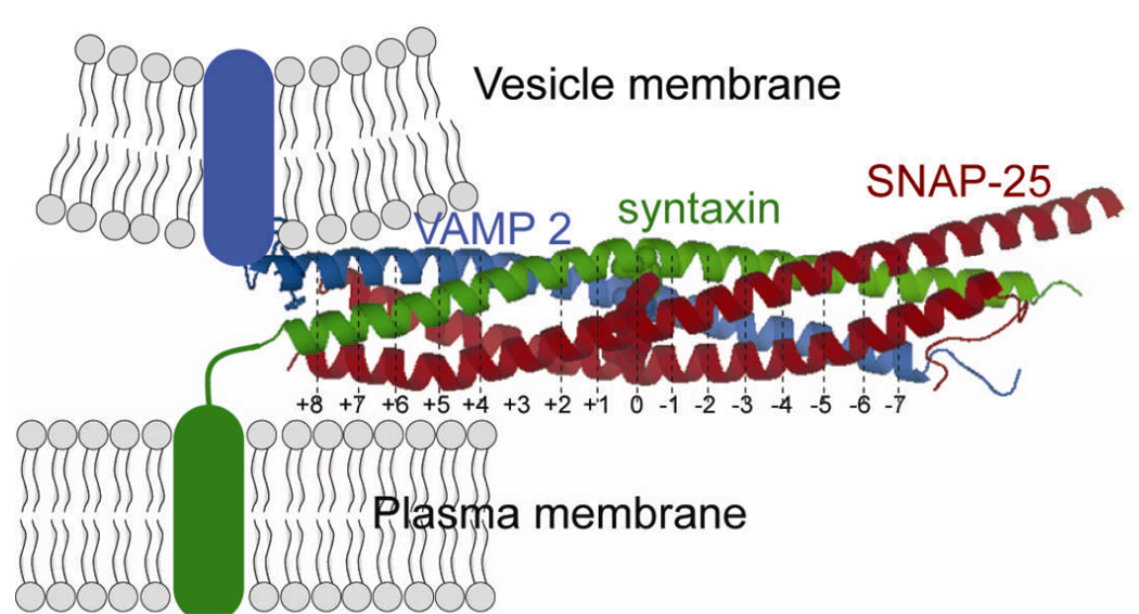
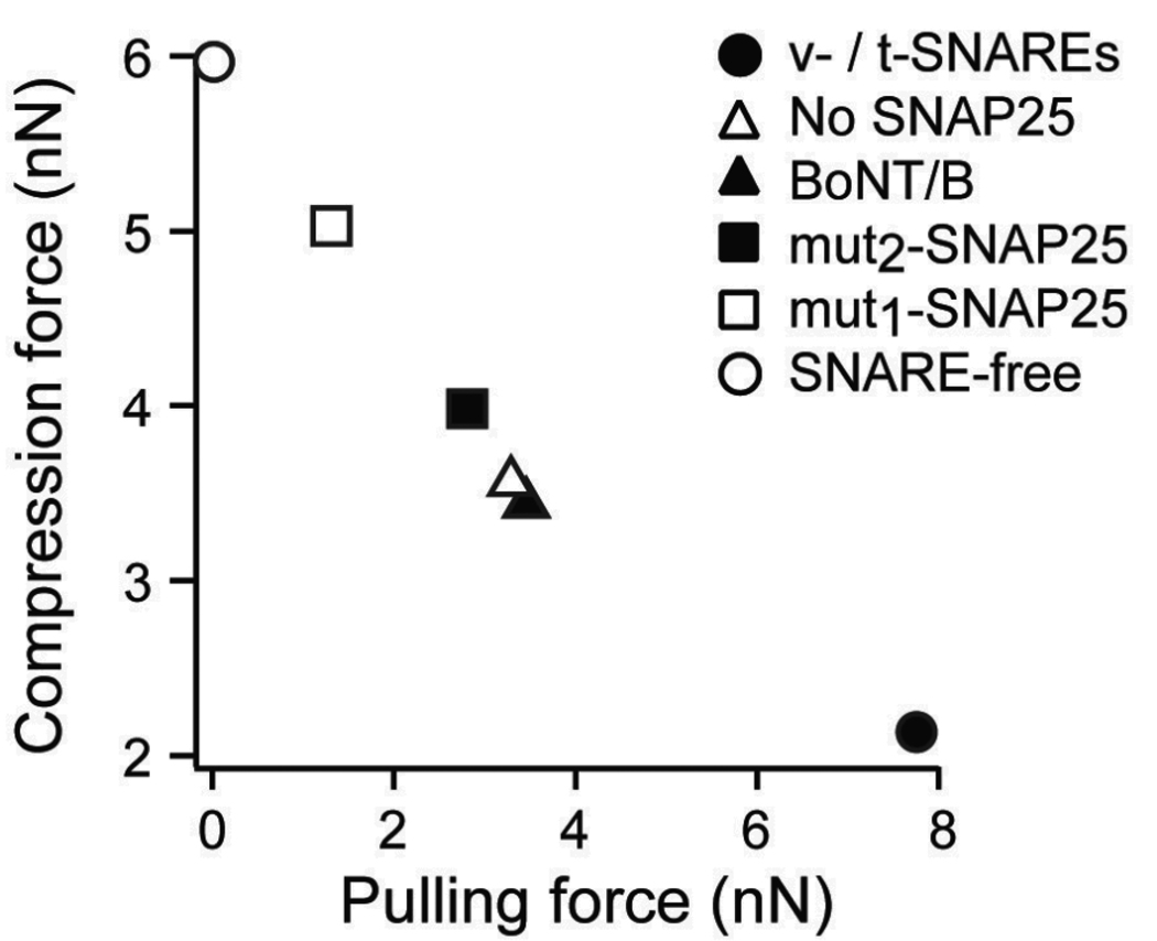
Membrane fusion is driven by specialized proteins that reduce the free energy penalty for the fusion process. In neurons and secretory cells, soluble N-ethylmaleimide-sensitive factor-attachment protein (SNAP) receptors (SNAREs) mediate vesicle fusion with the plasma membrane during vesicular content release. Although, SNAREs have been widely accepted as the minimal machinery for membrane fusion, the specific mechanism for SNARE-mediated membrane fusion remains an active area of research. Here, we summarize recent findings based on force measurements acquired in a novel experimental system that uses atomic force microscope (AFM) force spectroscopy to investigate the mechanism(s) of membrane fusion and the role of SNAREs in facilitating membrane hemifusion during SNARE-mediated fusion. In this system, protein-free and SNARE-reconstituted lipid bilayers are formed on opposite (trans) substrates and the forces required to induce membrane hemifusion and fusion or to unbind single v-/t-SNARE complexes are measured. The obtained results provide evidence for a mechanism by which the pulling force generated by interacting trans-SNAREs provides critical proximity between the membranes and destabilizes the bilayers at fusion sites by broadening the hemifusion energy barrier and consequently making the membranes more prone to fusion.


Figures

Fig. 1
Fig. 1
Vesicle fusion at the neuronal synapse. Molecular components of the neuronal fusion machinery include VAMP 2, SNAP-25, syntaxin, synaptotagmin and complexin. According to the SNARE hypothesis, synaptic vesicles fuse with the plasma membrane and release neurotransmitters upon triggering by action potentials.
Fig. 2
Fig. 2
Crystal structure of the synaptic ternary SNARE complex. VAMP 2 (blue) anchored in the vesicular membrane binds to the t-SNAREs syntaxin (green) and SNAP-25 (red). Syntaxin is anchored in the plasma membrane via a transmembrane domain, whereas, SNAP-25 is recruited via palmitoylation sites. SNAP-25 binds to syntaxin and VAMP 2 through the conserved sn1 and sn2 domains located in the N- and C-terminals, respectively. The interaction between v- and t-SNAREs forms a ternary core complex that has been described as a parallel α-helical bundle or a coiled coil structure. The SNARE complex is stabilized by 15 hydrophobic layers (+8, +7, …, −6, −7, but not 0)within the α-helical bundle. Residues (VAMP Arg56, syntaxin Gln226, sn1 Gln53, and sn2 Gln74) forming the ionic ‘0’ layer are shown in space-filled spheres.
Fig. 3
Fig. 3
Models for SNARE-mediated membrane fusion: (a) the proximity model and (b) the protein pore model.
Fig. 4
Fig. 4
AFM measurements of SNARE-mediated membrane fusion. (a) Photograph of AFM and of a microbead attached to the end of an AFM cantilever. (b) AFM compression force measurement (force vs. piezo displacement) of supported double lipid bilayers. At large separation, there is negligible interaction between the bilayers. Upon approach of the AFM cantilever, the bilayers are pressed against one another. With the continued application of force, the bilayers are compressed together until hemifusion takes place and a first jump (J1) is observed at ~ 450 pN (f1). The piezo-electric transducer displacement (d; insert) during the jump is a measure of its distance and reflects the thickness of the fused bilayer. A transient reduction in force takes place as the cantilever tip relaxes during the jump, followed by the continued application of compression, which eventually leads to the appearance of a second jump (J2) at ~ 900 pN (f2), which is indicative of the full fusion of the bilayers. On the other hand, during retraction of the cantilever the force decreases to zero as the cantilever returns to its relaxed position. No adhesion is observed in the presence of protein-free bilayers. The force measurements were performed in Tris buffered saline.
Fig. 5
Fig. 5
(a) Dynamic force spectra for the hemifusion of DMPC floating bilayers at different temperatures. (b) Dynamic force spectra for the hemifusion of egg PC floating bilayers with different cholesterol concentration. Lines are fits of the dynamic compression model described in the text to the data points. Error bars are the standard error of the mean (s.e.m.) all the measured compression forces required to induce hemifusion at the corresponding compression rates.
Fig. 6
Fig. 6
(a) Schematic illustration of the effect of applied force on the activation potential of the lipid bilayer hemifusion/fusion. The applied force adds a linear term (dotted line) to the thermopotential of the system, which effectively tilts the barrier and reduces the activation potential of the process. (b)Schematic depiction (not to scale) of the energy landscape for the hemifusion of DMPC floating bilayers below and above the melting temperature (Tm) of DMPC. During compression of the apposed individual floating bilayers the bilayers pass (along the gray dashed or dotted lines) through a transition state that is at the peak of an energy barrier. Arbitrary positions along the free energy axis were chosen for the unfused and hemifused bilayers since it is not known which of the states has the lower free energy minimum. On the other hand, we use the initial unfused state, where the compressed individual floating bilayers still exist, as a reference point along the reaction coordinate. Recall that no significant change in the barrier height was observed with temperature for DMPC floating bilayers (see Table 2); in contrast, a pronounced widening (0.61 Å) in the barrier was observed when DMPC bilayers were heated above Tm to 30°C. Broadening of the hemifusion barrier results in reducing its slope and consequently less energy input to overcome it which translates into a facilitation of the fusion process. (c) Kinetic profiles of DMPC membrane hemifusion above (dotted line) and below (solid line) Tm. The increased kinetics of hemifusion for the DMPC membranes is evident in accelerated fusion under compression.
Fig. 7
Fig. 7
(a) Dynamic force spectra of the hemifusion process for egg PC bilayers with and without SNAREs. The compression force required to induce hemifusion is significantly reduced when v- and t-SNAREs are incorporated in the opposite bilayers, respectively. Interaction between the opposite v- and t-SNAREs is inhibited by the cytoplasmic domain of VAMP (cd-VAMP) and consequently the observed reduction in the compression force is abolished. (b) Similarly, truncating SNAP-25 by mutations or VAMP by BoNT/B interfered with the SNARE interaction and increased the compression force required to induce hemifusion of egg PC bilayers. Lines are fits of eq. (1) to the data points. Error bars are the s.e.m.
Fig. 8
Fig. 8
(a) Energy landscape of SNARE-mediated membrane fusion. The combined effects of the SNAREs on the hemifusion activation barrier width and height result in a significant reduction in of energy requirements for membrane fusion when compared to SNARE-free bilayers. (b) The SNARE-mediated facilitation in the membrane hemifusion is evident in the kinetic profiles for egg PC bilayers with SNAREs (dotted line) and without SNAREs (solid line); a 4 fold increase in the hemifusion rate is initially observed in the absence of applied conpression and the rate increases exponentially under accelerated conditions with applied force.
Fig. 9
Fig. 9
Dynamic strength of the SNARE complex. (a) AFM force scan showing the force vs. piezo-displacement curve between apposed floating lipid bilayers containing SNAREs. The forced unbinding of the SNARE complex formed the v- and t-SNAREs in the opposite bilayers occurs during retraction of the AFM cantilever away from the substrate. fu is the unbinding force of the SNARE complex. (b) Dynamic force spectra for the unbinding of the SNARE complex with and without SNAP-25. An outer and an inner barrier activation barrier were revealed during the slow and fast loading regimes, respectively. The higher unbinding forces in the fast loading regime indicate a strong binding interaction of the native v-/t-SNARE complex due to the steeper inner barrier. (c) This is evident in the energy landscape for the unbinding of the SNARE complex with and without SNAP-25. Omission of SNAP-25 from the SNARE complex widened the inner activation barrier and resulted in reducing its slope and, consequently, a weaker interaction.
Fig. 10
Fig. 10
Membrane fusion facilitation quantified by the compression needed to induce hemifusion (fC) correlates with the binding force generated by interacting SNAREs as characterized by the SNARE pulling force (fπ)

Similar articles

Cited by

References

    1. Blumenthal R, Clague MJ, Durell SR, Epand RM. Chem. Rev. 2003;103:53. - PubMed
    1. Chernomordik LV, Kozlov MM. Annu. Rev. Biochem. 2003;72:175. - PubMed
    1. Fardon NJ, Wilkinson R, Thomas TH. Am. J. Hypertension. 2001;14:927. - PubMed
    1. Lentsch AB, Ward PA. J. Pathol. 2000;190:343. - PubMed
    1. Weber T, Zemelman BV, McNew JA, Westermann B, Gmachl M, Parlati F, Sollner TH, Rothman JE. Cell. 1998;92:759. - PubMed

LinkOut - more resources