. 2009 Dec 27;2(1):28-42.
doi: 10.18632/aging.100111.

Attainment of polarity promotes growth factor secretion by retinal pigment epithelial cells: relevance to age-related macular degeneration

Affiliations

Attainment of polarity promotes growth factor secretion by retinal pigment epithelial cells: relevance to age-related macular degeneration

Shozo Sonoda et al. Aging (Albany NY). .

Abstract

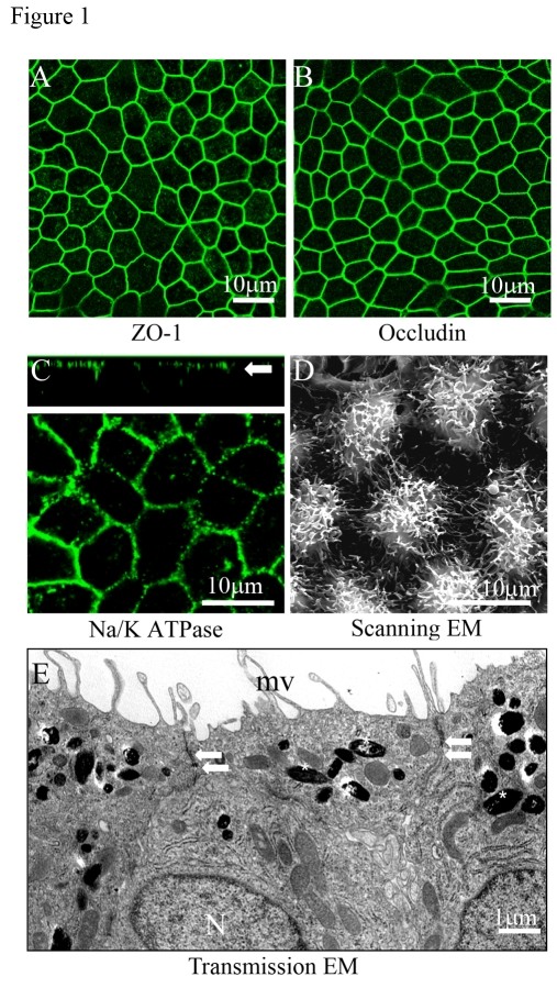
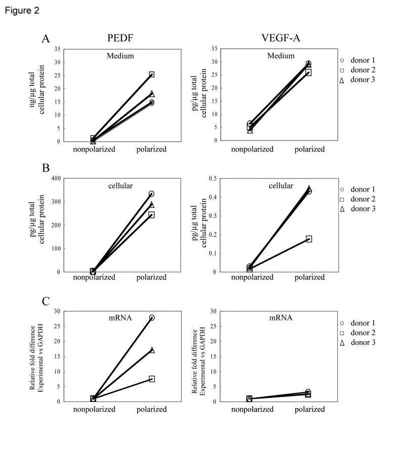
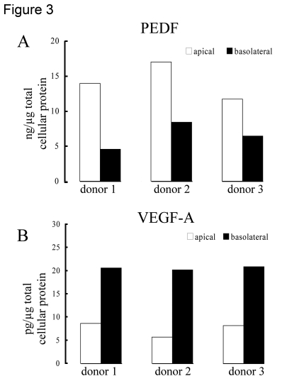
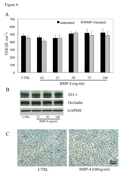
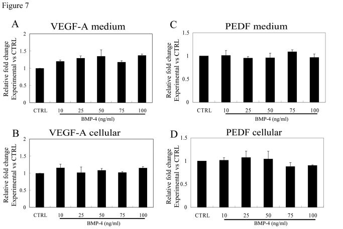
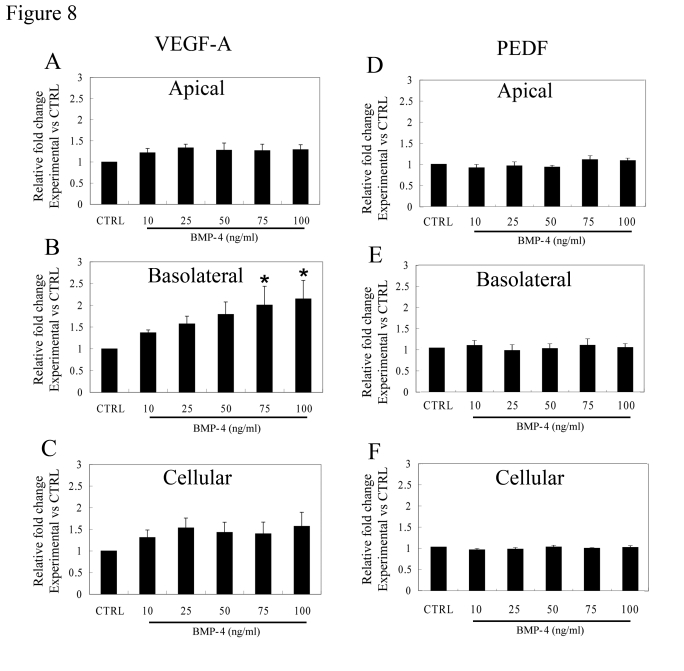
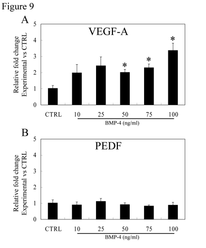
The antiangiogenic and neurotrophic growth factor, pigment epithelial derived factor (PEDF), and the proangiogenic growth factor, vascular endothelial growth factor-A (VEGF), are released from retinal pigment epithelial (RPE) cells where they play a critical role in the pathogenesis of age-related macular degeneration (AMD). Since RPE polarity may be altered in advanced AMD, we studied the effect of polarization of differentiated, human RPE monolayer cultures on expression and secretion of PEDF and VEGF. Polarized RPE demonstrated apical microvilli, expression of tight junction proteins, apical localization of Na/K- ATPase, and high transepithelial resistance (490 +/- 17 Omega x cm(2)). PEDF secretion was about 1000 fold greater than that for VEGF in both polarized and non-polarized cultures. Polarization of the RPE monolayer increased PEDF secretion, which was predominantly apical, by 34 fold (p<0.02) and VEGF secretion, which was predominantly basolateral, by 5.7 fold (p<0.02). Treatment of non-polarized RPE cultures with bone morphogenetic protein-4 (BMP-4) had no effect on PEDF or VEGF secretion, but resulted in a dose-dependent >2-fold increase in basolateral VEGF secretion (p<0.05) in polarized cultures. Our data show that polarity is an important determinant of the level of PEDF and VEGF secretion in RPE and support the contention that loss of polarity of RPE in AMD results in marked loss of neurotrophic and vascular support for the retina potentially leading to photoreceptor loss and blindness.

Keywords: BMP-4; PEDF; VEGF-A; age-related macular degeneration; cell polarity; retinal pigment epithelial cell.


Conflict of interest statement

The authors have no conflict of interests to declare.

Figures

Figure 1.
Figure 1.. Confocal and electron microscopic characterization of polarized RPE cells.
Evidence for tight junction proteins and polarity in fetal RPE cells cultured on Transwell filters for 6 weeks. (A, B) Immunofluorescence staining of tight junction proteins ZO-1 and occludin. (C) Localization of Na/K- ATPase to the apical plasma membrane as shown in the confocal vertical (X-Z) section (white arrow). (D) Well differentiated apical microvilli observed by scanning electron microscopy (SEM). (E) Well developed microvilli (mv), localization of pigment on the apical side (asterisks), nuclei on basal side (N), and presence of tight-junctional complexes (arrows) by transmission electron microscopy (TEM).
Figure 2.
Figure 2.. Differences in PEDF and VEGF secretion between nonpolarized and polarized RPE from various donors after 24h.
Secretion from the polarized RPE cells represent the sum of experimentally determined apical and basolateral secretion values, normalized for total cellular protein. The total secretion increased 34 fold for PEDF and 5.7 fold for VEGF-A (A). Analysis of cellular protein (B) and mRNA (C) showed that expression in polarized human RPE was higher compared to nonpolarized RPE cells.
Figure 3.
Figure 3.. Polarized secretion of PEDF and VEGF in differentiated human RPE cells.
Human polarized RPE cells on transwells isolated from 3 different donors preferentially secreted PEDF (A) to the apical side of the tissue and VEGF-A (B) to the basolateral side. The bars represent average of 2 determinations for each donor with variation in each sample <5%.
Figure 4.
Figure 4.. Distribution of PEDF in apical, central and basal regions in nonpolarized and polarized RPE cells by confocal microscopy.
Staining for PEDF is more intense in polarized RPE as compared to nonpolarized RPE. The apical region shows much higher PEDF expression in polarized cells.
Figure 5.
Figure 5.. Cell cycle analysis of polarized RPE monolayers.
(A, B, C) Expression of p27 (green) and its localization to nuclei (blue). (D, E, F). Polarized RPE cultures show lack of expression of Ki-67 (green) in the nuclei. Nuclei counterstained blue with DAPI.
Figure 6.
Figure 6.. Effect of BMP-4 treatment in highly differenti-ated RPE monolayers.
(A) Transepithelial resistance (TER) of polarized human RPE monolayers and effect of rhBMP-4 treatment. TER values in human RPE monolayers, maintained for 1 month in 1% FBS-containing medium, averaged 490 ±17 Ω. cm2 (mean ± SEM, n=48). The TER measurements in polarized RPE cells exposed to rhBMP-4 treatment for 24 h showed no significant difference (P>0.05) versus untreated controls (n=9/group). (B) Expression levels of tight junction proteins, ZO-1 and occludin were not significantly different between the BMP-4 treated and the untreated control groups. (C) No significant cell death was observed by TUNEL staining in highly polarized RPE cells of both untreated control and 100ng/ml BMP-4 treatment groups.
Figure 7.
Figure 7.. Effect of rhBMP-4 treatment on secretion of VEGF-A and PEDF from nonpolarized RPE cells.
Secretion of VEGF-A (A) and PEDF (C) are presented along with the corresponding cellular VEGF-A (B) and cellular PEDF (D) from three different donors. Data are presented as fold difference as compared to untreated controls. The cellular concentrations of VEGF-A and PEDF did not differ from untreated controls for the entire BMP-4 concentration range.
Figure 8.
Figure 8.. Effect of BMP-4 on VEGF-A and PEDF secretion from polarized RPE.
Fold change over control values calculated from ELISA analysis is presented to account for inter donor variations. (A) The increase in VEGF-A secretion after treatment with BMP-4 from the apical domain was not statistically significant (p>0.05). (B) An increase in VEGF-A secretion from the basolateral domain was found even with the lowest dose used (10ng/ml) which increased further in a dose-dependent fashion. Asterisk indicates that VEGF-A secretion with 75 and 100ng/ml BMP-4 treatment was significantly higher than that of control (p<0.05). (C) The cellular levels of VEGF-A were not significantly affected by BMP-4 treatment. (D, E, F) No significant change was observed for PEDF secretion either at the apical domain or the basolateral domain and in cellular PEDF levels. Data are mean±SEM from four different donors.
Figure 9.
Figure 9.. Effect of BMP-4 treatment on gene expression of VEGF-A and PEDF in polarized RPE.
Expression of VEGF-A (A) and PEDF (B) mRNA in polarized fetal RPE cells vs controls was analyzed by real-time PCR. BMP-4 treatment caused an increase in VEGF-A gene expression, especially at 50, 75, and 100ng/ml BMP-4 treatment which was significantly different from controls (p<0.05). PEDF mRNA did not change with BMP-4 dose for the BMP-4 dose range studied.

References

    1. Hogan MJ. Role of the retinal pigment epithelium in macular disease. Trans Am Acad Ophthalmol Otolaryngol. 1972;76:64–80. - PubMed
    1. Sheedlo HJ, Li L, Turner JE. Effects of RPE-cell factors secreted from permselective fibers on retinal cells in vitro. Brain Res. 1992;587:327–337. - PubMed
    1. Sonoda S, Spee C, Barron E, Ryan SJ, Kannan R, Hinton DR. A protocol for the culture and differentiation of highly polarized human retinal pigment epithelial cells. Nature Protocols. 2009;4:662–673. - PMC - PubMed
    1. Hu J, Bok D. A cell culture medium that supports the differentiation of human retinal pigment epithelium into functionally polarized monolayers. Mol Vis. 2001;7:14–19. - PubMed
    1. Holtkamp GM, Van Rossem M, de Vos AF, Willekens B, Peek R, Kijlstra A. Polarized secretion of IL-6 and IL-8 by human retinal pigment epithelial cells. Clin Exp Immunol. 1998;112:34–43. - PMC - PubMed

Publication types

MeSH terms