. 2009 Dec 10;2(1):43-62.
doi: 10.18632/aging.100107.

Local IGF-1 isoform protects cardiomyocytes from hypertrophic and oxidative stresses via SirT1 activity

Affiliations

Local IGF-1 isoform protects cardiomyocytes from hypertrophic and oxidative stresses via SirT1 activity

Manlio Vinciguerra et al. Aging (Albany NY). .

Abstract

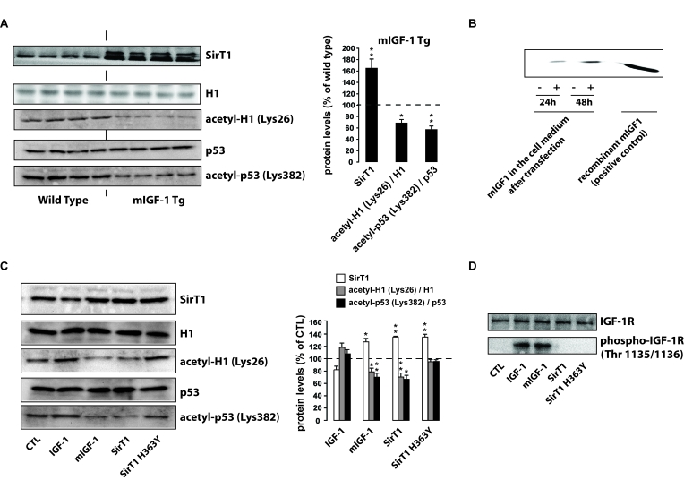
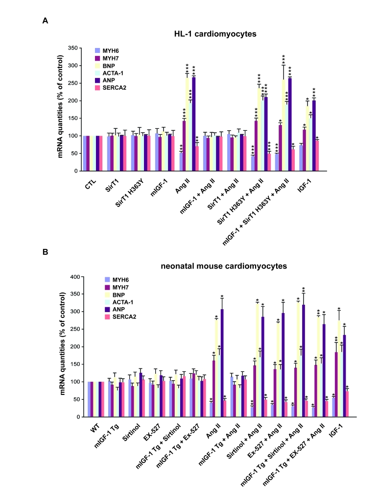
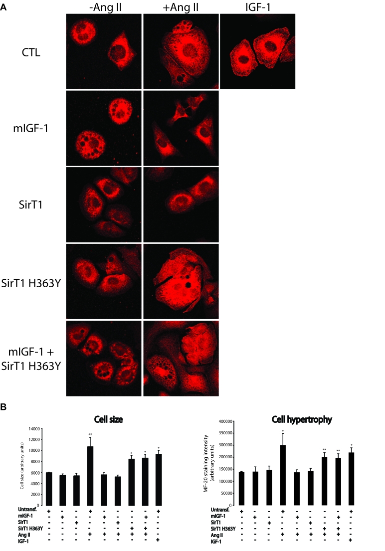
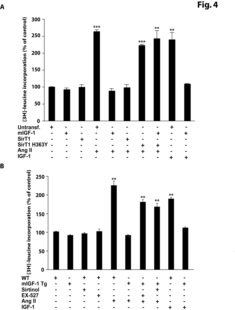
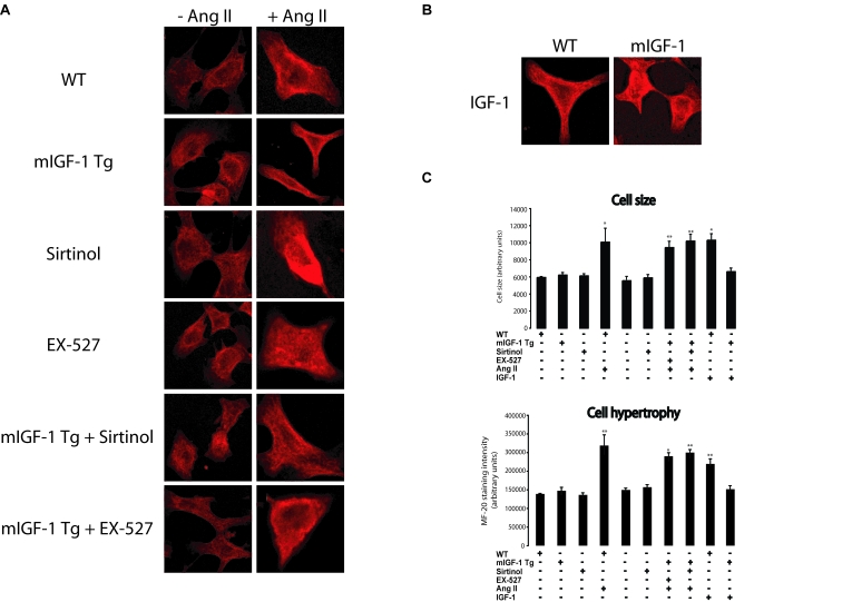
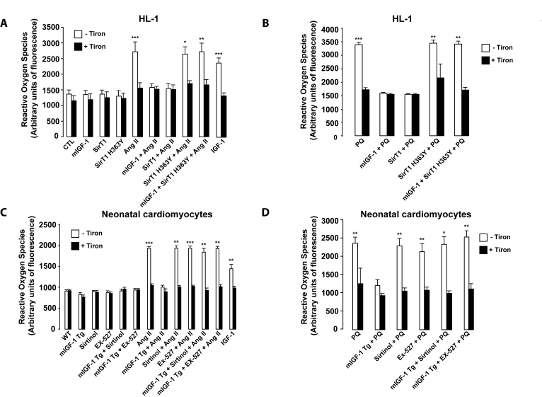
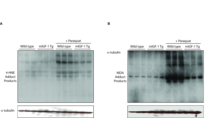
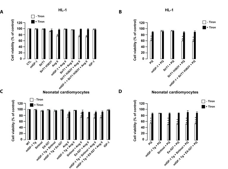
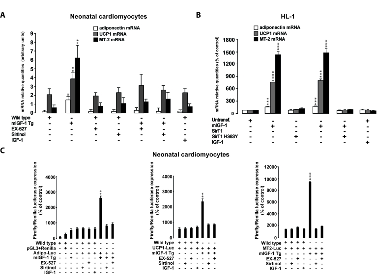
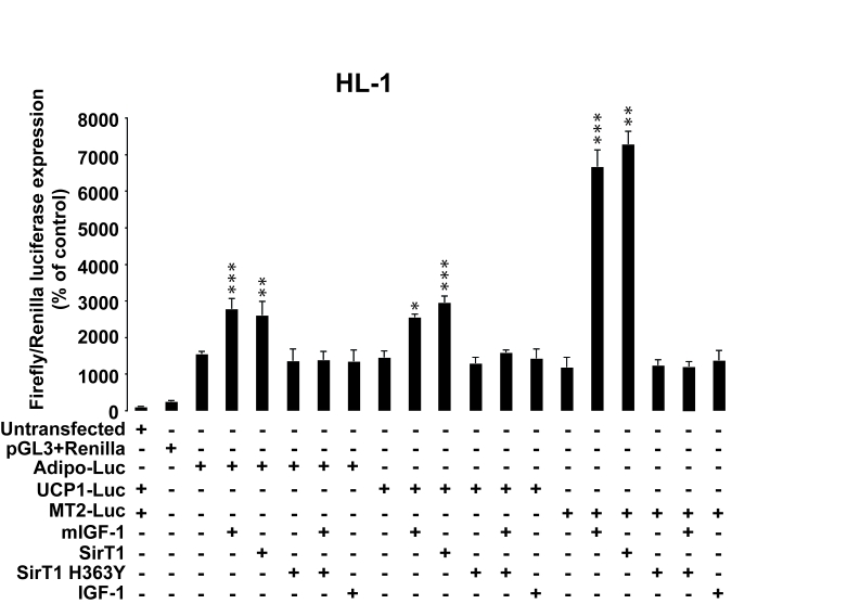
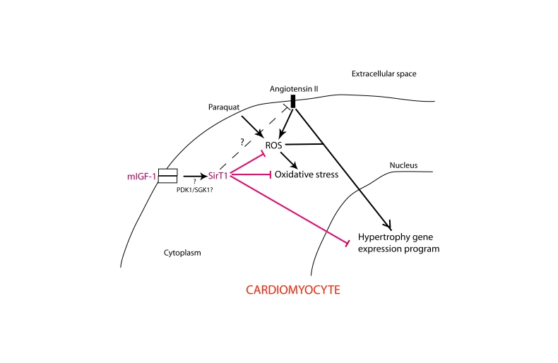
Oxidative and hypertrophic stresses contribute to the pathogenesis of heart failure. Insulin-like growth factor-1 (IGF-1) is a peptide hormone with a complex post-transcriptional regulation, generating distinct isoforms. Locally acting IGF-1 isoform (mIGF-1) helps the heart to recover from toxic injury and from infarct. In the murine heart, moderate overexpression of the NAD(+)-dependent deacetylase SirT1 was reported to mitigate oxidative stress. SirT1 is known to promote lifespan extension and to protect from metabolic challenges. Circulating IGF-1 and SirT1 play antagonizing biological roles and share molecular targets in the heart, in turn affecting cardiomyocyte physiology. However, how different IGF-1 isoforms may impact SirT1 and affect cardiomyocyte function is unknown. Here we show that locally acting mIGF-1 increases SirT1 expression/activity, whereas circulating IGF-1 isoform does not affect it, in cultured HL-1 and neonatal cardiomyocytes. mIGF-1-induced SirT1 activity exerts protection against angiotensin II (Ang II)-triggered hypertrophy and against paraquat (PQ) and Ang II-induced oxidative stress. Conversely, circulating IGF-1 triggered itself oxidative stress and cardiomyocyte hypertrophy. Interestingly, potent cardio-protective genes (adiponectin, UCP-1 and MT-2) were increased specifically in mIGF-1-overexpressing cardiomyocytes, in a SirT1-dependent fashion. Thus, mIGF-1 protects cardiomyocytes from oxidative and hypertrophic stresses via SirT1 activity, and may represent a promising cardiac therapeutic.

Keywords: IGF-1; SirT1; cardiomyocytes; cell hypertrophy; oxidative stress.


Conflict of interest statement

The authors of this article report no conflict of interests.

Figures

Figure 1.
Figure 1.. mIGF-1, but not IGF-1, increases SirT1 expression and activity in mouse cardiomyocytes.
(A) Left panel: representative Western blots of SirT1, histone H1, acetyl-H1 (Lys26), detected in nuclear extracts, and of p53 and acetyl-p53 (Lys382), detected in whole tissue lysates, from wild type and mIGF-1 Tg mice. Four animals of a total of 10 are shown; right panel: densitometric quantification of SirT1, acetyl-H1(Lys26)/H1 and acetyl-p53(Lys382)/p53 levels in cardiomyocytes from mIGF-1 mice, expressed as % of those in wild type cardiomyocytes. (B) representative Western Blot of mIGF-1 detected in the extracellular medium of HL-1 cardiomyocytes, transfected with a plasmid carrying mouse mIGF-1 cDNA. (C) Left panel: representative Western Blot of SirT1, histone H1, acetyl-H1 (Lys26) detected in nuclear extracts, and of p53 and acetyl-p53 (Lys382) detected in whole cell lysates, from HL-1 cardiomyocytes transfected with the indicated constructs (SirT1 or SirT1 H363Y) or treated with 20 ng/ml IGF-1 for 24 hours; right panel: densitometric quantification of SirT1, acetyl-H1(Lys26)/H1 and acetyl-p53(Lys382)/p53 levels in transfected or treated cells, expressed as % of control (CTL). (D) Representative Western blots of IGF-1 receptor (IGF-1R) or phospho-IGF-1R (on Thr 1135/1136) in HL-1 cardiomyocytes lysates. Results in (A) and (B) are means ± SE of 3 independent experiments (**,***p versus unstimulated control cells or untreated WT cardiomyocytes).
Figure 2.
Figure 2.. mIGF-1 prevents Ang II- and IGF-1-induced fetal gene program activation.
(A) HL-1 cardiomyocytes were transfected with the indicated plasmids, or treated with 20 ng/ml IGF-1 for 24 h, before exposure to Ang II (1 μM for 24 h). Untransfected cells were used as control (CTL). (B) Neonatal mouse cardiomyocytes from wild type (WT) or heart overexpressing mIGF-1 mice (mIGF-1 Tg) were pre-incubated with sirtinol (100 μM) or EX-527 (1 μM), or treated with 20 ng/ml IGF-1 for 24 h, prior to exposure to Ang II (1 μM for 24 h). Untreated WT cardiomyocytes were used as control. (A, B) The expression levels of MYH6, MYH7, BNP, ACTA-1, ANP and SERCA2 mRNAs were examined by qRT-PCR. Results are means ± SE of 3 independent experiments (*,**,***p versus unstimulated control cells).
Figure 3.
Figure 3.. mIGF-1 prevents Ang II- and IGF-1-induced cell hypertrophy (MF-20 staining) in HL-1 cardiomyocytes.
(A) HL-1 cardiomyocytes were transfected or treated as in Legend of Figure 2A. Sarcomeric myosin was stained with MF-20 antibody and images were acquired using a Leica confocal microscope. (B) Cell size and cell hypertrophy quantified according to MF-20 staining in HL-1 cardiomyocytes in the different experimental conditions as in as in Legend of Figure 2A. Results are means ± SE of 3 independent experiments (*,**p versus unstimulated control cells). Bar: 25 μM.
Figure 4.
Figure 4.. mIGF-1 prevents Ang II- and IGF-1-induced cell hypertrophy ([3H]-leucine incorporation) in HL-1 cardiomyocytes and in mouse neonatal primary cardiomyocytes.
(A) HL-1 cardiomyocytes were transfected with the indicated plasmids, or treated with 20 ng/ml IGF-1 for 24 h, or exposed to Ang II (1 μM for 24 h). Untransfected cells were used as control (CTL). Together with Ang II, HL-1 cells were also incubated with 1μCi/ml of [3H]-labeled leucine (24 h). (B) Neonatal primary cardiomyocytes from wild type or mIGF-1 Tg mice were treated with SirT1 inhibitors (sirtinol, 100 μM; EX-527, 1 μM), or treated with 20 ng/ml IGF-1 for 24 h, or exposed to Ang II (1 μM for 24 h); concomitantly to Ang II addition, cells were incubated with 1mCi/ml of [3H]-labeled leucine (24 h). (A, B) [3H]-leucine incorporation values were normalized to total protein content and expressed as % of control. Results are means ± SE of 3 independent experiments (**,***p versus unstimulated control cells or untreated WT cardiomyocytes).
Figure 5.
Figure 5.. mIGF-1 prevents Ang II- and IGF-1-induced cell hypertrophy (MF-20 staining) in mouse neonatal primary cardiomyocytes.
(A) Neonatal primary cardiomyocytes from wild type or mIGF-1 Tg mice were treated as in Legend of Figure 2B. (B) Cell size and hypertrophy were quantified according to MF-20 staining in the different experimental conditions as in as in Legend of Figure 2B. Results are means ± SE of 3 independent experiments (*,**p versus unstimulated control cells). Bar: 25 μM.
Figure 6.
Figure 6.. mIGF-1 prevents Ang II-, PQ- and IGF-1-induced increase in reactive oxygen species (ROS) generation in HL-1 cardiomyocytes and in mouse neonatal primary cardiomyocytes.
(A, B) HL-1 cardiomyocytes were transfected or treated as in Legend of Figure 2A, except that Ang II (1 μM) or PQ (100 μM) were added for only 60 min. Untransfected cells were used as control (CTL). (C, D) Neonatal primary cardiomyocytes from wild type or mIGF-1 Tg mice were treated as in Legend of Figure 2A, except that Ang II (1 μM) or PQ (100 μM) were added for only 60 min. (A-D) ROS production was monitored with the fluorescent probe dichlorofluorescein diacetate (CM-DCFDA) and fluorescence values were normalized to protein content. Results are means ± SE of 3 independent experiments (*,**,***p versus unstimulated control cells or untreated WT cardiomyocytes).
Figure 7.
Figure 7.. mIGF-1 protects the murine heart from PQ-induced oxidative stress.
PQ was injected intraperitoneally at a concentration of 30 mg/kg, while control animals were injected with a saline solution. All mice were sacrificed 24 hours after injections. The figure shows representative Western blots of 4-hydroxy-2-nonenal (4-HNE) adduct products (left panel) and of malondialdehyde (MDA) adduct products (right panel) from wild type, mIGF-1 Tg, wild type plus PQ and mIGF-1 mice plus PQ. Three animals of a total of 10 are shown in both panels A and B.
Figure 8.
Figure 8.. mIGF-1 prevents Ang II- and PQ-dependent cell death in HL-1 cardiomyocytes and in mouse neonatal primary cardiomyocytes.
(A, B) HL-1 cardiomyocytes were transfected or treated as in Legend of Figure 2A. (C, D) Neonatal primary cardiomyocytes from wild type or mIGF-1 Tg mice were treated as in Legend of Figure 2B. (A-D) Cell viability was monitored with propidium iodide (PI) by flow cytometry and values were normalized to protein content. Results are means ± SE of 3 independent experiments (*,**,***p versus unstimulated control cells or untreated wild type cardiomyocytes).
Figure 9.
Figure 9.. SirT1 is necessary for mIGF-1-dependent upregulation of anti-oxidant and hypertrophic genes adiponectin, UCP1 and MT-2.
(A) Neonatal primary cardiomyocytes from wild type or mIGF-1 Tg mice were treated with sirtinol (100 μM) or EX-527 (1 μM), or treated with 20 ng/ml IGF-1 for 24 h. (B) HL-1 cardiomyocytes were transfected with the indicated plasmids, or treated with 20 ng/ml IGF-1 for 24 h. Untransfected cells were used as control (CTL). (A, B) The expression levels of adiponectin, UCP-1 and MT-2 mRNAs were examined by Real Time-PCR. (C) Neonatal primary cardiomyocytes from wild type or mIGF-1 Tg mice, and HL-1 cardiomyocytes, were transfected with 1 μg of plasmids carrying Firefly luciferase under the control of promoters of adiponectin (Adipo-Luc), UCP1 (UCP1-Luc) and MT-2 (MT-2-Luc) genes, respectively, together with 1 μg of Renilla Luciferase plasmid. Neonatal primary cardiomyocytes were also treated with different inhibitors or IGF-1 as described in (A). Dual luciferase assays were performed in duplicate for each condition. (A-C) Results are means ± SE of 3 independent experiments (*,**,***p versus untreated cardiomyocytes).
Figure 10.
Figure 10.. SirT1 is necessary for promoter-dependent mIGF-1-dependent upregulation of anti-oxidant and hypertrophic genes adiponectin, UCP1 and MT-2.
HL-1 cardiomyocytes were transfected with the indicated plasmids, and/or treated with 20 ng/ml IGF-1 for 24 h. HL-1 cardiomyocytes were also co-transfected with 1 μg of plasmids carrying Firefly luciferase under the control of promoters of adiponectin (Adipo-Luc), UCP1 (UCP1-Luc) and MT-2 (MT-2-Luc) genes, respectively, together with 1 μg of Renilla Luciferase plasmid. Untransfected cells were used as control. Dual luciferase assays were performed in duplicate for each condition. Results are means ± SE of 3 independent experiments (*,**,***p versus untransfected/unstimulated control cells).
Figure 11.
Figure 11.. Simplified scheme illustrating the role of mIGF-1-induced SirT1 activity in protection against Ang II- and PQ-mediated oxidative stress and hypertrophy in cardiomyocytes.
Question point and dashed line indicate unanswered issues and hypothetical signaling cross-talk, respectively.

Comment in

  • Surprising sirtuin crosstalk in the heart.
    Schug TT, Li X. Schug TT, et al. Aging (Albany NY). 2010 Mar 31;2(3):129-32. doi: 10.18632/aging.100128. Aging (Albany NY). 2010. PMID: 20375467 Free PMC article. No abstract available.

Similar articles

Cited by

References

    1. Frey N, Olson EN. Cardiac hypertrophy: the good, the bad, and the ugly. Annu Rev Physiol. 2003;65:45–79. - PubMed
    1. Giordano FJ. Oxygen, oxidative stress, hypoxia, and heart failure. J Clin Invest. 2005;115:500–508. - PMC - PubMed
    1. Takimoto E, Kass DA. Role of oxidative stress in cardiac hypertrophy and remodeling. Hypertension. 2007;49:241–248. - PubMed
    1. Seddon M, Looi YH, Shah AM. Oxidative stress and redox signalling in cardiac hypertrophy and heart failure. Heart. 2007;93:903–907. - PMC - PubMed
    1. Lavu S, Boss O, Elliott PJ, Lambert PD. Sirtuins--novel therapeutic targets to treat age-associated diseases. Nat Rev Drug Discov. 2008;7:841–853. - PubMed

Publication types

MeSH terms

LinkOut - more resources