

Signaling From Lysosomes Enhances Mitochondria-Mediated Photodynamic Therapy In Cancer Cells
- PMID: 20228965
- PMCID: PMC2836728
- DOI: 10.1117/12.823752
Signaling From Lysosomes Enhances Mitochondria-Mediated Photodynamic Therapy In Cancer Cells
Abstract
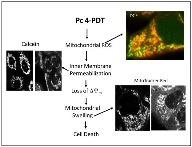
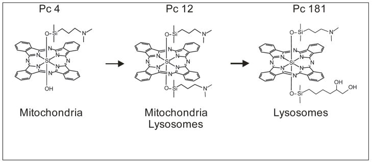
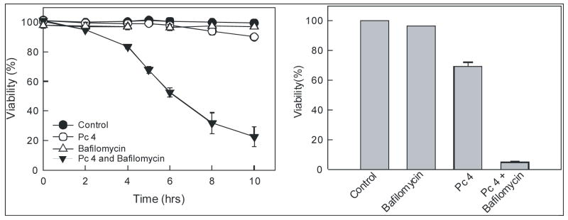
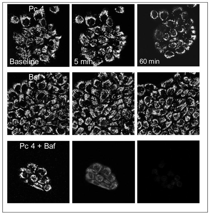
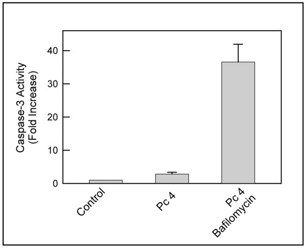
In photodynamic therapy (PDT), visible light activates a photosensitizing drug added to a tissue, resulting in singlet oxygen formation and cell death. Assessed by confocal microscopy, the photosensitizer phthalocyanine 4 (Pc 4) localizes primarily to mitochondrial membranes in cancer cells, resulting in mitochondria-mediated cell death. A Pc 4 derivative, Pc 181, accumulates into lysosomes. In comparison to Pc 4, Pc 181 was a more effective photosensitizer promoting killing cancer cells after PDT. The mode of cell death after Pc 181-PDT is predominantly apoptosis, and pancaspase and caspase-3 inhibitors prevent onset of the cell death. To assess further how lysosomes contribute to PDT, we monitored cell killing of A431cells after PDT in the presence and absence of bafilomycin, an inhibitor of the acidic vacuolar proton pump that collapses the pH gradient of the lysosomal/endosomal compartment. Bafilomycin by itself did not induce toxicity but greatly enhanced Pc 4-PDT-induced cell killing. In comparison to Pc 4, less enhancement of cell killing by bafilomycin occurred after Pc 181-PDT at photosensitizer doses producing equivalent cell killing in the absence of bafilomycin. These results indicate that lysosomal disruption can augment PDT with Pc 4, which targets predominantly mitochondria, but less so after PDT with Pc 181, since Pc 181 already targets lysosomes.
Figures
Similar articles
-
Lysosomal signaling enhances mitochondria-mediated photodynamic therapy in A431 cancer cells: role of iron.Photochem Photobiol. 2012 Mar-Apr;88(2):461-8. doi: 10.1111/j.1751-1097.2012.01081.x. Epub 2012 Jan 25. Photochem Photobiol. 2012. PMID: 22220628 Free PMC article.
-
Mitoferrin-2-dependent mitochondrial iron uptake sensitizes human head and neck squamous carcinoma cells to photodynamic therapy.J Biol Chem. 2013 Jan 4;288(1):677-86. doi: 10.1074/jbc.M112.422667. Epub 2012 Nov 7. J Biol Chem. 2013. PMID: 23135267 Free PMC article.
-
The peripheral benzodiazepine receptor in photodynamic therapy with the phthalocyanine photosensitizer Pc 4.Photochem Photobiol. 2002 Jun;75(6):652-61. doi: 10.1562/0031-8655(2002)075<0652:tpbrip>2.0.co;2. Photochem Photobiol. 2002. PMID: 12081328
-
Signaling pathways in cell death and survival after photodynamic therapy.J Photochem Photobiol B. 2000 Aug;57(1):1-13. doi: 10.1016/s1011-1344(00)00065-8. J Photochem Photobiol B. 2000. PMID: 11100832 Review.
-
Singlet Oxygen, Photodynamic Therapy, and Mechanisms of Cancer Cell Death.J Oncol. 2022 Jun 25;2022:7211485. doi: 10.1155/2022/7211485. eCollection 2022. J Oncol. 2022. PMID: 35794980 Free PMC article. Review.
Cited by
-
Intracellular detection of singlet oxygen using fluorescent nanosensors.Analyst. 2021 Jun 14;146(12):3933-3941. doi: 10.1039/d1an00456e. Analyst. 2021. PMID: 33982697 Free PMC article.
-
Effects of photodynamic treatment on mesenchymal stromal cells.Dokl Biol Sci. 2013 May;450:185-8. doi: 10.1134/S0012496613030174. Epub 2013 Jul 3. Dokl Biol Sci. 2013. PMID: 23821063 No abstract available.
-
Typical and Atypical Inducers of Lysosomal Cell Death: A Promising Anticancer Strategy.Int J Mol Sci. 2018 Aug 1;19(8):2256. doi: 10.3390/ijms19082256. Int J Mol Sci. 2018. PMID: 30071644 Free PMC article. Review.
-
Cyanine dyes in the mitochondria-targeting photodynamic and photothermal therapy.Commun Chem. 2024 Aug 13;7(1):180. doi: 10.1038/s42004-024-01256-6. Commun Chem. 2024. PMID: 39138299 Free PMC article. Review.
-
Effect of Curcumin-Loaded Mesoporous Silica Nanoparticles on the Head and Neck Cancer Cell Line, HN5.Curr Issues Mol Biol. 2022 Oct 27;44(11):5247-5259. doi: 10.3390/cimb44110357. Curr Issues Mol Biol. 2022. PMID: 36354669 Free PMC article.
References
-
- Oleinick NL, Morris RL, Belichenko I. The role of apoptosis in response to photodynamic therapy: what, where, why, and how. Photochem Photobiol Sci. 2002;1:1–21. - PubMed
-
- Lam M, Oleinick NL, Nieminen AL. Photodynamic therapy-induced apoptosis in epidermoid carcinoma cells. Reactive oxygen species and mitochondrial inner membrane permeabilization. J Biol Chem. 2001;276:47379–47386. - PubMed
-
- Trivedi NS, Wang HW, Nieminen AL, Oleinick NL, Izatt JA. Quantitative analysis of Pc 4 localization in mouse lymphoma (LY-R) cells via double-label confocal fluorescence microscopy. Photochem Photobiol. 2000;71:634–639. - PubMed
-
- Oleinick NL, Antunez AR, Clay ME, Rihter BD, Kenney ME. New phthalocyanine photosensitizers for photodynamic therapy. Photochem Photobiol. 1993;57:242–247. - PubMed
Grants and funding
LinkOut - more resources
Full Text Sources
Other Literature Sources
Research Materials