. 2010 Aug;248(8):1105-16.
doi: 10.1007/s00417-010-1336-7. Epub 2010 Mar 13.

Exogenous modulation of intrinsic optic nerve neuroprotective activity

Affiliations

Exogenous modulation of intrinsic optic nerve neuroprotective activity

Sinisa D Grozdanic et al. Graefes Arch Clin Exp Ophthalmol. 2010 Aug.

Abstract

Background: To characterize the molecular and functional status of the rat retina and optic nerve after acute elevation of intraocular pressure (IOP).

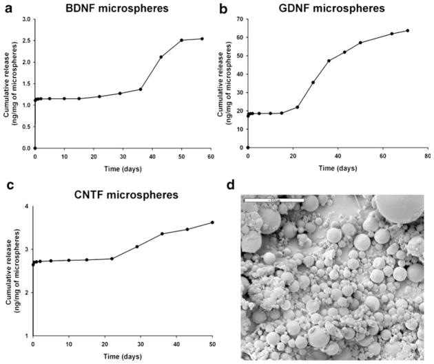
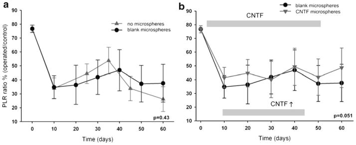
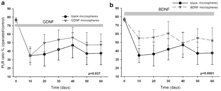
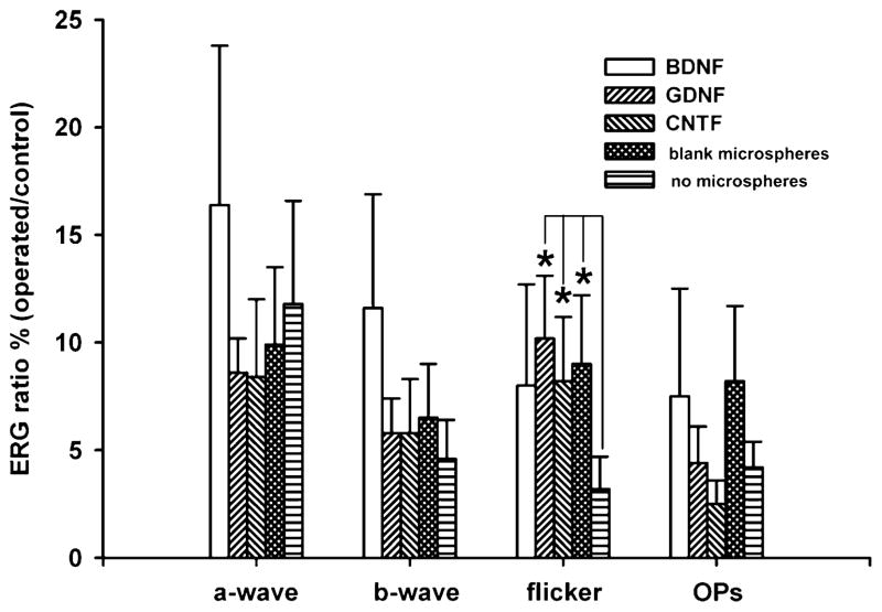
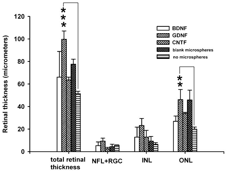
Methods: Retinal ischemia was induced in rats by increasing the IOP (110 mmHg/60 minutes). Microarray analysis, quantitative RT-PCR (qRT-PCR) and immunohistochemistry were used to characterize retinal tissue. PLGA microspheres containing neurotrophic factors (BDNF, GDNF, or CNTF) or empty microspheres were injected into the vitreous of operated animals 1 day after elevation of IOP. Pupil light reflex (PLR) parameters and electroretinograms (ERG) were monitored at multiple time points during the 60-day postoperative recovery period.

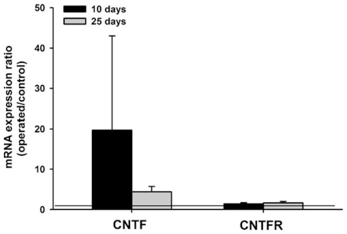
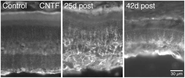
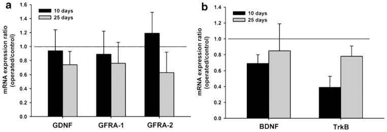
Results: Molecular analysis showed a significant intrinsic up-regulation of CNTF at 10 and 25 days after induction of the acute ocular hypertension (p = 0.0067). Molecular tissue analysis of GDNF and its receptors (GDNFR1, GDNFR2), and BDNF and its receptor (trkB) showed no change in expression. Animals that received CNTF microspheres had no significant functional recovery compared to animals which received blank microspheres (p > 0.05). Animals that received GDNF or BDNF microspheres showed significant PLR recovery (p < 0.05 and p < 0.001 respectively) compared to non-treated animals.

Conclusions: Continuous release of neurotrophic growth factors (NGFs) significantly protects optic nerve function in the experimental model of retinal ischemia observed by PLR analysis.


Figures

Fig. 1
Fig. 1
Release kinetics of PLGA microspheres (a, b, c)—steady release of neurotrophic growth factors was detected for at least 60 days (a, BDNF), 80 days (b, GDNF) and 50 days (c, CNTF). Scanning electron microscopy of the microspheres (d). Scale bar in D=50 μm
Fig. 2
Fig. 2
Real-time PCR analysis showed significant CNTF mRNA up-regulation in operated eyes 10 and 25 days postoperatively (horizontal line represents ratio value 1, which corresponds to equal expression of mRNA in operated and control eyes). Analysis of CNTF receptor (CNTFR) mRNA levels did not reveal significant change between ischemic and control eyes (bars represent mean+SD)
Fig. 3
Fig. 3
Immunohistochemical analysis showed increased CNTF protein expression at 25 days postoperatively (time interval of spontaneous functional recovery). However, protein expression declined at 42 days postoperatively, which corresponded to the decline in PLR and ERG function in operated animals. Despite intrinsic growth factor production, significant inner retina thinning is present at 42 days postoperatively
Fig. 4
Fig. 4
a Real-time PCR analysis showed relatively unchanged mRNA expression for GDNF and its respective receptors (GFRA1 and 2) 10 and 25 days post ischemic insult (horizontal line represents ratio value of 1, which corresponds to equal expression of mRNA in operated and control eyes). b Analysis of the BDNF and its respective receptor TrkB, showed a trend toward decreased expression at 10 days; however, mRNA levels normalized 25 days post ischemic insult (bars represent mean+SD)
Fig. 5
Fig. 5
a Graph shows pupil light reflex data from rodent eyes exposed to ischemic insult that received blank microspheres. There was no significant difference between the 2 groups. b Graph shows the group that received CNTF microspheres. While overall function in CNTF treated rats was somewhat better, difference was not statistically significant when compared to rats which received empty microspheres. Values are plotted as mean±SEM
Fig. 6
Fig. 6
a GDNF microspheres provided significant recovery of PLR function compared to control blank microspheres, starting 10 days post ischemic insult. b BDNF showed an immediate positive effect on retinal function which was sustained until the end of the experiment. Values are plotted as mean±SEM
Fig. 7
Fig. 7
Electroretinography analysis revealed significantly better cone function in those operated eyes that received GDNF, CNTF or blank microspheres than in eyes which had retinal ischemic episode but did not receive any intraocular injection (bars represent standard error of mean, * represents p<0.05, OPs=oscillatory potentials). Bars represent mean+SEM
Fig. 8
Fig. 8
Morphometric analysis showed significant preservation of retinal structure in the GDNF-treated group when compared to animals which did not receive intraocular injection. However, difference was not significant when data were compared with ischemic eyes which received blank microspheres (bars represent standard error of mean, ** represents p<0.01, *** represents p<0.0001). Bars represent mean±SEM

Similar articles

Cited by

References

    1. Adachi M, Takahashi K, Nishikawa M, Miki H, Uyama M. High intraocular pressure-induced ischemia and reperfusion injury in the optic nerve and retina in rats. Graefes Arch Clin Exp Ophthalmol. 1996;234:445–451. - PubMed
    1. Hughes WF. Quantitation of ischemic damage in the rat retina. Exp Eye Res. 1991;53:573–582. - PubMed
    1. Quigley HA. Neuronal death in glaucoma. Prog Retin Eye Res. 1999;18:39–57. - PubMed
    1. Takahashi H, Goto T, Shoji T, Tanito M, Park M, Chihara E. Diabetes-associated retinal nerve fiber damage evaluated with scanning laser polarimetry. Am J Ophthalmol. 2006;142:88–94. - PubMed
    1. Ko ML, Hu DN, Ritch R, Sharma SC. The combined effect of brain-derived neurotrophic factor and a free radical scavenger in experimental glaucoma. Invest Ophthalmol Vis Sci. 2000;41:2967–2971. - PubMed

Publication types

MeSH terms

Substances