

Sox10 is necessary for oligodendrocyte survival following axon wrapping
- PMID: 20229602
- PMCID: PMC3639140
- DOI: 10.1002/glia.20981
Sox10 is necessary for oligodendrocyte survival following axon wrapping
Abstract
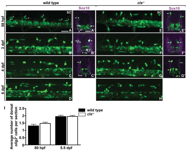
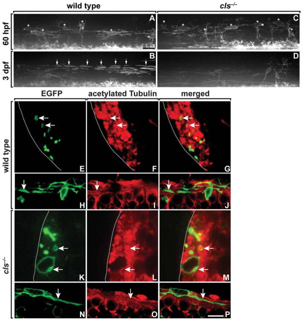
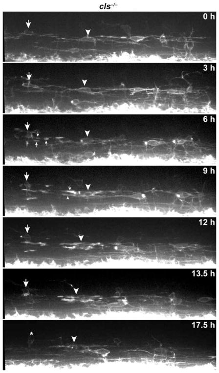
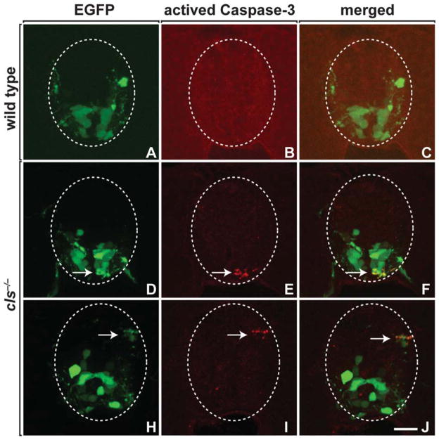
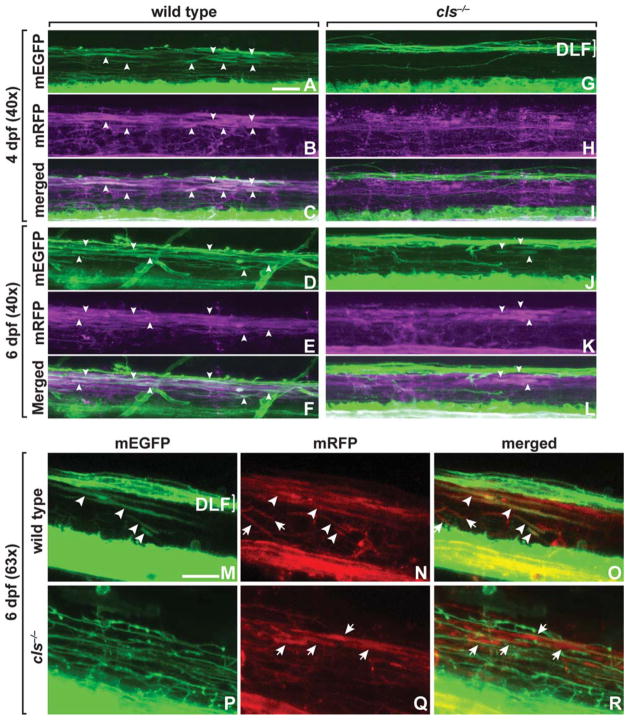
Cells of the oligodendrocyte lineage, which form the myelinating glia of the vertebrate central nervous system, undergo a stepwise developmental progression entailing specification from neuroepithelial precursors, proliferation, migration to expand and distribute the population, and differentiation to ensheath axons with myelin. Understanding the genetic mechanisms that regulate each of these steps during development is important, because this might lead to therapies to promote remyelination following neural injury or disease. Genetic studies in mice indicated that the Sox10 transcription factor is required during the differentiation stage to promote myelin gene expression. However, whether Sox10 also promotes other features of oligodendroctye differentiation remained unknown. In this study, we used time-lapse imaging to investigate the behavior and fates of oligodendrocyte lineage cells in zebrafish embryos and larvae that lacked Sox10 function. This revealed that the myelinating subset of oligodendrocyte progenitor cells (OPCs) migrates, divides, and wraps axons normally, but then dies. Nonmyelinating oligodendrocyte progenitors divided more frequently, maintaining a normal population size. New oligodendrocytes produced by these progenitors wrapped axons and survived, but did not express myelin genes at high levels. We conclude that, in addition to promoting myelin gene expression, Sox10 function is necessary for the survival of myelinating oligodedrocytes subsequent to axon wrapping but is not required for the survival of nonmyelinating OPCs.
Figures


Similar articles
-
nkx2.2a promotes specification and differentiation of a myelinating subset of oligodendrocyte lineage cells in zebrafish.Neuron Glia Biol. 2008 May;4(2):71-81. doi: 10.1017/S1740925X09990123. Neuron Glia Biol. 2008. PMID: 19737431 Free PMC article.
-
Olig2+ precursors produce abducens motor neurons and oligodendrocytes in the zebrafish hindbrain.J Neurosci. 2009 Feb 25;29(8):2322-33. doi: 10.1523/JNEUROSCI.3755-08.2009. J Neurosci. 2009. PMID: 19244509 Free PMC article.
-
Knockdown of Lingo1b protein promotes myelination and oligodendrocyte differentiation in zebrafish.Exp Neurol. 2014 Jan;251:72-83. doi: 10.1016/j.expneurol.2013.11.012. Epub 2013 Nov 18. Exp Neurol. 2014. PMID: 24262204
-
New insights into signaling during myelination in zebrafish.Curr Top Dev Biol. 2011;97:1-19. doi: 10.1016/B978-0-12-385975-4.00007-3. Curr Top Dev Biol. 2011. PMID: 22074600 Free PMC article. Review.
-
Epigenetic control of oligodendrocyte development: adding new players to old keepers.Curr Opin Neurobiol. 2016 Aug;39:133-8. doi: 10.1016/j.conb.2016.06.002. Epub 2016 Jun 14. Curr Opin Neurobiol. 2016. PMID: 27308779 Free PMC article. Review.
Cited by
-
Developmental exposure to domoic acid targets reticulospinal neurons and leads to aberrant myelination in the spinal cord.Sci Rep. 2023 Feb 14;13(1):2587. doi: 10.1038/s41598-023-28166-2. Sci Rep. 2023. PMID: 36788234 Free PMC article.
-
The visual system of zebrafish and its use to model human ocular diseases.Dev Neurobiol. 2012 Mar;72(3):302-27. doi: 10.1002/dneu.20919. Dev Neurobiol. 2012. PMID: 21595048 Free PMC article. Review.
-
Myelin gene regulatory factor is required for maintenance of myelin and mature oligodendrocyte identity in the adult CNS.J Neurosci. 2012 Sep 5;32(36):12528-42. doi: 10.1523/JNEUROSCI.1069-12.2012. J Neurosci. 2012. PMID: 22956843 Free PMC article.
-
pyPAGE: A framework for Addressing biases in gene-set enrichment analysis-A case study on Alzheimer's disease.PLoS Comput Biol. 2024 Sep 5;20(9):e1012346. doi: 10.1371/journal.pcbi.1012346. eCollection 2024 Sep. PLoS Comput Biol. 2024. PMID: 39236079 Free PMC article.
-
A principled strategy for mapping enhancers to genes.Sci Rep. 2019 Jul 30;9(1):11043. doi: 10.1038/s41598-019-47521-w. Sci Rep. 2019. PMID: 31363138 Free PMC article.
References
-
- Barres BA, Jacobson MD, Schmid R, Sendtner M, Raff MC. Does oligodendrocyte survival depend on axons? Curr Biol. 1993;3:489–497. - PubMed
-
- Baumann N, Pham-Dinh D. Biology of oligodendrocyte and myelin in the mammalian central nervous system. Physiol Rev. 2001;81:871–927. - PubMed
-
- Benninger Y, Colognato H, Thurnherr T, Franklin RJ, Leone DP, Atanasoski S, Nave KA, Ffrench-Constant C, Suter U, Relvas JB. β1-Integrin signaling mediates premyelinating oligodendrocyte survival but is not required for CNS myelination and remyelination. J Neurosci. 2006;26:7665–7673. - PMC - PubMed
-
- Brosamle C, Halpern ME. Characterization of myelination in the developing zebrafish. Glia. 2002;39:47–57. - PubMed
-
- Butt AM, Hornby MF, Ibrahim M, Kirvell S, Graham A, Berry M. PDGF-α receptor and myelin basic protein mRNAs are not coexpressed by oligodendrocytes in vivo: A double in situ hybridization study in the anterior medullary velum of the neonatal rat. Mol Cell Neurosci. 1997;8:311–322. - PubMed
Publication types
MeSH terms
Substances
Grants and funding
LinkOut - more resources
Full Text Sources
Molecular Biology Databases
