

Comparison of organ doses for patients undergoing balloon brachytherapy of the breast with HDR 192Ir or electronic sources using monte carlo simulations in a heterogeneous human phantom
- PMID: 20229875
- PMCID: PMC2905452
- DOI: 10.1118/1.3292292
Comparison of organ doses for patients undergoing balloon brachytherapy of the breast with HDR 192Ir or electronic sources using monte carlo simulations in a heterogeneous human phantom
Abstract
Purpose: Accelerated partial breast irradiation via interstitial balloon brachytherapy is a fast and effective treatment method for certain early stage breast cancers. The radiation can be delivered using a conventional high-dose rate (HDR) 192Ir gamma-emitting source or a novel electronic brachytherapy (eBx) source which uses lower energy x rays that do not penetrate as far within the patient. A previous study [A. Dickler, M. C. Kirk, N. Seif, K. Griem, K. Dowlatshahi, D. Francescatti, and R. A. Abrams, "A dosimetric comparison of MammoSite high-dose-rate brachytherapy and Xoft Axxent electronic brachytherapy," Brachytherapy 6, 164-168 (2007)] showed that the target dose is similar for HDR 192Ir and eBx. This study compares these sources based on the dose received by healthy organs and tissues away from the treatment site.
Methods: A virtual patient with left breast cancer was represented by a whole-body, tissue-heterogeneous female voxel phantom. Monte Carlo methods were used to calculate the dose to healthy organs in a virtual patient undergoing balloon brachytherapy of the left breast with HDR 192Ir or eBx sources. The dose-volume histograms for a few organs which received large doses were also calculated. Additional simulations were performed with all tissues in the phantom defined as water to study the effect of tissue inhomogeneities.
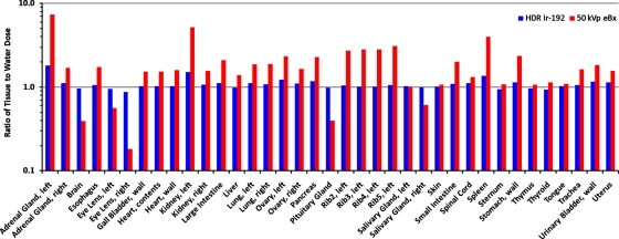
Results: For both HDR 192Ir and eBx, the largest mean organ doses were received by the ribs, thymus gland, left lung, heart, and sternum which were close to the brachytherapy source in the left breast, eBx yielded mean healthy organ doses that were more than a factor of approximately 1.4 smaller than for HDR 192Ir for all organs considered, except for the three closest ribs. Excluding these ribs, the average and median dose-reduction factors were approximately 28 and approximately 11, respectively. The volume distribution of doses in nearby soft tissue organs that were outside the PTV were also improved with eBx. However, the maximum dose to the closest rib with the eBx source was 5.4 times greater than that of the HDR 192Ir source. The ratio of tissue-to-water maximum rib dose for the eBx source was approximately 5.
Conclusions: The results of this study indicate that eBx may offer lower toxicity to most healthy tissues, except nearby bone. TG-43 methods have a tendency to underestimate dose to bone, especially the ribs. Clinical studies evaluating the negative health effects caused by irradiating healthy organs are needed so that physicians can better understand when HDR 192Ir or eBx might best benefit a patient.
Figures


Similar articles
-
A dosimetric comparison of 169Yb and 192Ir for HDR brachytherapy of the breast, accounting for the effect of finite patient dimensions and tissue inhomogeneities.Med Phys. 2006 Dec;33(12):4583-9. doi: 10.1118/1.2392408. Med Phys. 2006. PMID: 17278810
-
Comparison of tumor and normal tissue dose for accelerated partial breast irradiation using an electronic brachytherapy eBx source and an Iridium-192 source.J Appl Clin Med Phys. 2010 Sep 14;11(4):3301. doi: 10.1120/jacmp.v11i4.3301. J Appl Clin Med Phys. 2010. PMID: 21081891 Free PMC article.
-
Calculated organ doses using Monte Carlo simulations in a reference male phantom undergoing HDR brachytherapy applied to localized prostate carcinoma.Med Phys. 2013 Mar;40(3):033901. doi: 10.1118/1.4791647. Med Phys. 2013. PMID: 23464344
-
Accelerated partial breast irradiation: a dosimetric comparison of three different techniques.Brachytherapy. 2005;4(2):121-9. doi: 10.1016/j.brachy.2004.12.005. Brachytherapy. 2005. PMID: 15893265
-
Xoft Axxent electronic brachytherapy: a new device for delivering brachytherapy to the breast.Nat Clin Pract Oncol. 2009 Mar;6(3):138-42. doi: 10.1038/ncponc1319. Epub 2009 Jan 27. Nat Clin Pract Oncol. 2009. PMID: 19174776 Review.
Cited by
-
Electronic brachytherapy as adjuvant therapy for early stage breast cancer: a retrospective analysis.Onco Targets Ther. 2011 Jan 12;4:13-20. doi: 10.2147/OTT.S15297. Onco Targets Ther. 2011. PMID: 21552411 Free PMC article.
-
Incorporation of Electronic Brachytherapy for Skin Cancer into a Community Dermatology Practice.J Clin Aesthet Dermatol. 2015 Nov;8(11):28-32. J Clin Aesthet Dermatol. 2015. PMID: 26705437 Free PMC article.
-
Isolating the impact of tissue heterogeneities in high dose rate brachytherapy treatment of the breast.Phys Imaging Radiat Oncol. 2025 Feb 22;33:100737. doi: 10.1016/j.phro.2025.100737. eCollection 2025 Jan. Phys Imaging Radiat Oncol. 2025. PMID: 40093657 Free PMC article.
-
Radiotherapy of early-stage breast cancer.Precis Radiat Oncol. 2023 Jan 29;7(1):67-79. doi: 10.1002/pro6.1183. eCollection 2023 Mar. Precis Radiat Oncol. 2023. PMID: 40336616 Free PMC article. Review.
-
Computed organ doses to an Indian reference adult during brachytherapy treatment of esophagus, breast, and neck cancers.J Med Phys. 2012 Jul;37(3):151-4. doi: 10.4103/0971-6203.99238. J Med Phys. 2012. PMID: 22973082 Free PMC article.
References
-
- Belkacémi Y., Hannoun-Lévi J. M., and Lartigau E., in New Technologies in Radiation Oncology, edited by Schlegel W., Bortfeld T., and Grosu A. -L. (Springer, New York, 2006), pp. 397–407.10.1007/3-540-29999-8_31 - DOI
-
- Smith B. D., Arthur D. W., Buchholz T. A., Haffty B. G., Hahn C. A., Hardenbergh P. H., Julian T. B., Marks L. B., Todor D. A., Vicini F. A., Whelan T. J., White J., Wo J. Y., and Harris J. R., “Accelerated partial breast irradiation consensus statement from the American society for radiation oncology (ASTRO),” Int. J. Radiat. Oncol., Biol., Phys. IOBPD3 74, 987–1001 (2009). - PubMed
-
- Stewart A. J., O’Farrell D. A., Cormack R. A., Hansen J. L., Khan A. J., Mutyala S., and Devlin P. M., “Dose volume histogram analysis of normal structures associated with accelerated partial breast irradiation delivered by high dose rate brachytherapy and comparison with whole breast external beam radiotherapy fields,” Radiat. Oncol. 3(39) (2008).10.1186/1748-717X-3-39 - DOI - PMC - PubMed
-
- Adkison J. B., Kuske R. R., Quiet C. A., Beriwal S., and Patel R. R., “Five-year results of accelerated partial breast irradiation for ductal carcinoma in situ treated by interstitial multicatheter high-dose-rate brachytherapy or MammoSite,” Brachytherapy BRACC4 8, 107–108 (2009).10.1016/j.brachy.2009.03.013 - DOI
-
- Adkison J. B., Patel R. R., Beriwal S., Quiet C. A., and Kuske R. R., “Five-year results for accelerated partial breast irradiation using MammoSite balloon brachytherapy: The first 100 patients,” Brachytherapy BRACC4 7, 104 (2008).10.1016/j.brachy.2008.02.371 - DOI
Publication types
MeSH terms
Grants and funding
LinkOut - more resources
Full Text Sources
Medical
Miscellaneous
