

Contribution of optical zone decentration and pupil dilation on the change of optical quality after myopic photorefractive keratectomy in a cat model
- PMID: 20229950
- PMCID: PMC2841296
- DOI: 10.3928/1081597X-20100224-04
Contribution of optical zone decentration and pupil dilation on the change of optical quality after myopic photorefractive keratectomy in a cat model
Abstract
Purpose: To simulate the simultaneous contribution of optical zone decentration and pupil dilation on retinal image quality using wavefront error data from a myopic photorefractive keratectomy (PRK) cat model.
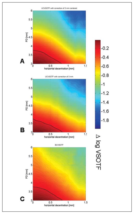
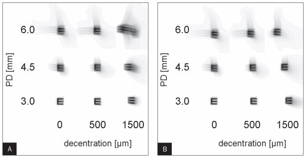
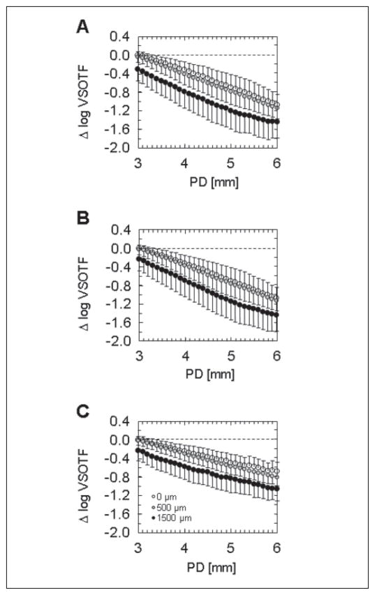
Methods: Wavefront error differences were obtained from five cat eyes 19+/-7 weeks (range: 12 to 24 weeks) after spherical myopic PRK for -6.00 diopters (D) (three eyes) and -10.00 D (two eyes). A computer model was used to simulate decentration of a 6-mm sub-aperture relative to the measured wavefront error difference. Changes in image quality (visual Strehl ratio based on the optical transfer function [VSOTF]) were computed for simulated decentrations from 0 to 1500 mum over pupil diameters of 3.5 to 6.0 mm in 0.5-mm steps. For each eye, a bivariate regression model was applied to calculate the simultaneous contribution of pupil dilation and decentration on the pre- to postoperative change of the log VSOTF.
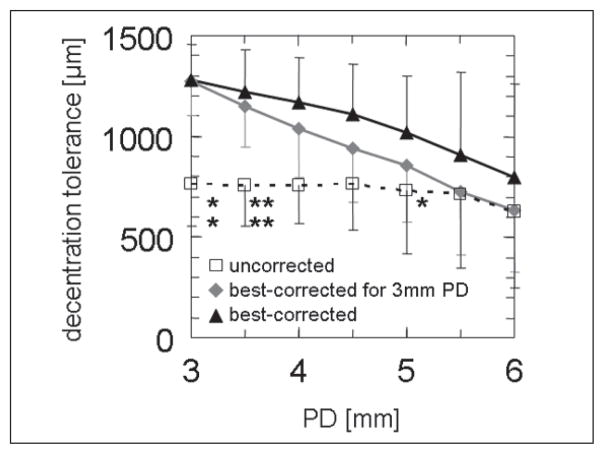
Results: Pupil diameter and decentration explained up to 95% of the variance of VSOTF change (adjusted R(2)=0.95). Pupil diameter had a higher impact on VSOTF (median beta=-0.88, P<.001) than decentration (median beta=-0.45, P<.001). If decentration-induced lower order aberrations were corrected, the impact of decentration further decreased (beta=-0.26) compared to the influence of pupil dilation (beta=-0.95).
Conclusions: Both pupil dilation and decentration of the optical zone affected the change of retinal image quality (VSOTF) after myopic PRK with decentration exerting a lower impact on VSOTF change. Thus, under physiological conditions pupil dilation is likely to have more effect on VSOTF change after PRK than optical zone decentration.
Copyright 2010, SLACK Incorporated.
Figures
References
-
- Seiler T, Kaemmerer M, Mierdel P, Krinke HE. Ocular optical aberrations after photorefractive keratectomy for myopia and myopic astigmatism. Arch Ophthalmol. 2000;118:17–21. - PubMed
-
- Moreno-Barriuso E, Lloves JM, Marcos S, Navarro R, Llorente L, Barbero S. Ocular aberrations before and after myopic corneal refractive surgery: LASIK-induced changes measured with laser ray tracing. Invest Ophthalmol Vis Sci. 2001;42:1396–1403. - PubMed
-
- Kohnen T, Bühren J, Kühne C, Mirshahi A. Wavefront-guided LASIK with the Zyoptix 3.1 system for the correction of myopia and compound myopic astigmatism with 1-year follow-up: clinical outcome and change in higher order aberrations. Ophthalmology. 2004;111:2175–2185. - PubMed
-
- Bühren J, Kohnen T. Factors affecting the change in lower-order and higher-order aberrations after wavefront-guided laser in situ keratomileusis for myopia with the Zyoptix 3.1 system. J Cataract Refract Surg. 2006;32:1166–1174. - PubMed
-
- Freedman KA, Brown SA, Mathews SM, Young RS. Pupil size and the ablation zone in laser refractive surgery: considerations based on geometric optics. J Cataract Refract Surg. 2003;29:1924–1931. - PubMed
Publication types
MeSH terms
Grants and funding
LinkOut - more resources
Full Text Sources
Miscellaneous
