. 2010 Apr 7;132(13):4815-21.
doi: 10.1021/ja910317a.

Sequential rearrangement of interhelical networks upon rhodopsin activation in membranes: the Meta II(a) conformational substate

Affiliations

Sequential rearrangement of interhelical networks upon rhodopsin activation in membranes: the Meta II(a) conformational substate

Ekaterina Zaitseva et al. J Am Chem Soc. .

Abstract

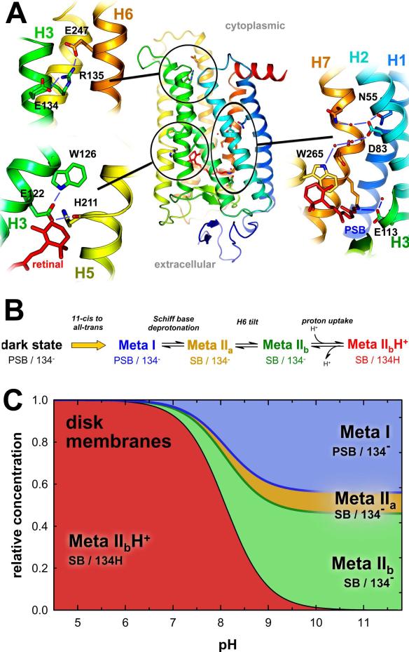
Photon absorption by rhodopsin is proposed to lead to an activation pathway that is described by the extended reaction scheme Meta I <==>Meta II(a) <==> Meta II(b) <==> Meta II(b)H(+), where Meta II(b)H(+) is thought to be the conformational substate that activates the G protein transducin. Here we test this extended scheme for rhodopsin in a membrane bilayer environment by investigating lipid perturbation of the activation mechanism. We found that symmetric membrane lipids having two unsaturated acyl chains, such as 1,2-dioleoyl-sn-glycero-3-phosphocholine (DOPC), selectively stabilize the Meta II(a) substate in the above mechanism. By combining FTIR and UV-visible difference spectroscopy, we characterized the structural and functional changes involved in the transition to the Meta II(a) intermediate, which links the inactive Meta I intermediate with the Meta II(b) states formed by helix rearrangement. Besides the opening of the Schiff base ionic lock, the Meta II(a) substate is characterized by an activation switch in a conserved water-mediated hydrogen-bonded network involving transmembrane helices H1/H2/H7, which is sensed by its key residue Asp83. On the other hand, movement of retinal toward H5 and its interaction with another interhelical H3/H5 network mediated by His211 and Glu122 is absent in Meta II(a). The latter rearrangement takes place only in the subsequent transition to Meta II(b), which has been previously associated with movement of H6. Our results imply that activating structural changes in the H1/H2/H7 network are triggered by disruption of the Schiff base salt bridge and occur prior to other chromophore-induced changes in the H3/H5 network and the outward tilt of H6 in the activation process.


Figures

Figure 1
Figure 1. Extended Meta I-Meta II scheme for rhodopsin activation explains visual function by an ensemble of conformational substates
(A) Activation of rhodopsin involves several conformational switches, salt bridges, and interhelical networks, of which several are highlighted here in the dark state of rhodopsin . (B) Triggering of these switches in the photoproducts of rhodopsin leads to a sequential progression of intermediates from inactive Meta I to the signaling state, involving formation of Meta IIa after disruption of the PSB salt bridge, Meta IIb after movement of H6, and Meta IIbH+ after proton uptake by Glu134, which were initially characterized only in a detergent environment . (C) In native disk membranes, the pH-independent thermodynamic equilibria between the Meta I, Meta IIa, and Meta IIb photoproduct states and the pH-dependent transition to Meta IIbH+ gives rise to complex pH-dependent population curves that can be determined by combination of FTIR and UV-visible spectrometries .
Figure 2
Figure 2. FTIR and UV-visible marker bands in pH-dependent experiments monitor conformation of the receptor and protonation state of the retinal chromophore
Light-induced FTIR difference spectra photoproduct minus dark state were obtained at a series of pH values at 30 °C with rhodopsin in disk membranes (A). The pH-dependent transition from pure Meta IIbH+ at acidic pH to a mixture of mostly Meta I and Meta IIb states at alkaline pH is shown (see Fig. 1C). In disk membranes, this transition affects all spectral features in a mostly concerted way, including carboxylic acid and amide I bands. In DOPC membranes (B), the pH-dependence is selectively lagging for the band of Asp83 at 1768 cm–1 (shown enlarged in the inset) and an unassigned band at 1687 cm–1. Corresponding UV-visible difference spectra (C and D) allow one to monitor the protonation status of the retinal Schiff base under identical conditions as in the FTIR experiments.
Figure 3
Figure 3. The Meta IIa conformational substate is selectively stabilized in DOPC membranes
The pH-dependence of FTIR marker bands of Figure 2 and of the protonation status of the retinal Schiff base (Θ=0 for fully protonated and Θ=1 for fully deprotonated, determined by UV-visible spectrometry) are compared for rhodopsin in disk membranes (A), in POPC membranes (B), and in DOPC membranes (C) (see text for details on the Θ values). While the curves obtained for disk and POPC membranes reveal only small differences in the alkaline endpoint value for the different markers, this does not hold for the curves obtained for DOPC membranes. Here the alkaline endpoint values of the Glu122 and amide marker bands are considerably lower than that of Schiff base deprotonation. This reflects substantial population of the Meta IIa conformational substate in DOPC membranes (D), which has a deprotonated Schiff base but lacks the H6 movement characteristic of the Meta IIb state. The changes sensed by Asp83, on the other hand, follow the same curve as retinal Schiff base protonation, indicating a triggering of the Asp83 activation switch already in Meta IIa.
Figure 4
Figure 4. Structural and functional transitions of rhodopsin intermediates
Representations of the Meta substates summarize results of complementary biophysical studies ,,,- and highlight structural and functional changes in the activation pathway of rhodopsin. This study adds essential structural information for the Meta IIa intermediate: opening of the PSB salt bridge is accompanied by activating conformational changes in the water-mediated H1/H2/H7 network around Asp83, while movement of retinal toward H5, which triggers the larger movements of H5 and H6, occurs only in the transition to Meta IIb.
None

Similar articles

Cited by

References

    1. Hofmann KP, Scheerer P, Hildebrand PW, Choe HW, Park JH, Heck M, Ernst OP. Trends Biochem. Sci. 2009;34:540–552. - PubMed
    1. Schwartz TW, Hubbell WL. Nature. 2008;455:473–474. - PMC - PubMed
    1. Ahuja S, Smith SO. Trends Pharmacol. Sci. 2009;30:494–502. - PubMed
    1. Oldham WM, Hamm HE. Nat. Rev. Mol. Cell Biol. 2008;9:60–71. - PubMed
    1. Lewis JW, Kliger DS. Methods Enzymol. 2000;315:164–178. - PubMed

Publication types