Review
. 2010 Jun;23(3):314-37.
doi: 10.1111/j.1755-148X.2010.00693.x. Epub 2010 Mar 8.

Genetic and environmental melanoma models in fish

Affiliations
Free PMC article
Review

Genetic and environmental melanoma models in fish

E Elizabeth Patton et al. Pigment Cell Melanoma Res. 2010 Jun.
Free PMC article

Abstract

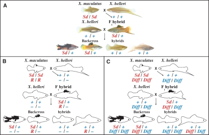
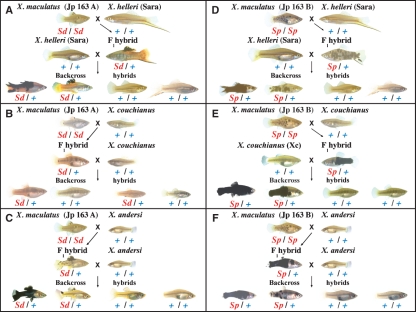
Experimental animal models are extremely valuable for the study of human diseases, especially those with underlying genetic components. The exploitation of various animal models, from fruitflies to mice, has led to major advances in our understanding of the etiologies of many diseases, including cancer. Cutaneous malignant melanoma is a form of cancer for which both environmental insult (i.e., UV) and hereditary predisposition are major causative factors. Fish melanoma models have been used in studies of both spontaneous and induced melanoma formation. Genetic hybrids between platyfish and swordtails, different species of the genus Xiphophorus, have been studied since the 1920s to identify genetic determinants of pigmentation and melanoma formation. Recently, transgenesis has been used to develop zebrafish and medaka models for melanoma research. This review will provide a historical perspective on the use of fish models in melanoma research, and an updated summary of current and prospective studies using these unique experimental systems.


Figures

Figure 1
Figure 1
Genetics of the Gordon-Kosswig spontaneous melanoma model. (A) Hybridization of the platyfish X . maculatus, exhibiting the macromelanophore spotted dorsal (Sd) pigment pattern, to the swordtail X. helleri generates F1 hybrids with an enhanced Sd pigment pattern on the dorsal fin. Backcrossing F1 hybrids to the X. helleri swordtail species generates first generation backcross hybrids (BC1 hybrids) with three phenotypes, as shown at the bottom of panel A. Approximately one-half of the BC1 hybrids are non-macromelanophore pigmented fish exhibiting no melanistic pigmentation (fish shown at lower right of panel A); these hybrids have not inherited the sex-linked Sd-Mdl allele (designated in the figure as Sd) from the original platyfish parent and therefore are not susceptible to melanoma. Of the remaining approximately one-half of BC1 hybrids, half of these (∼ 25% of total BC1 progeny) are heavily pigmented and develop invasive, exophytic, nodular malignant melanoma (lower left individual in panel A) and the other half (∼ 25% of BC1 progeny) show enhanced Sd pigmentation resembling the F1 hybrid phenotype, but only rarely develop melanoma late in life. (B) Hypothetical two-gene inheritance model explaining the apparently Mendelian inheritance of BC1 phenotypes. In this model, R is a platyfish gene that regulates the expression of the Xmrk oncogene associated with the Sd-Mdl allele, and its total loss in heavily pigmented BC1 hybrids that develop melanoma explains the melanoma susceptibility of these hybrids. Heterozygosity for R in lightly pigmented BC1 hybrids results in some regulation of Xmrk and inhibits melanoma formation. (C) Alternative two-gene inheritance model. In this model, the autosomal locus Diff regulates melanoma susceptibility but is not restricted to the platyfish parent, instead existing as alleles in Xiphophorus spp. populations. Mendelian inheritance of melanoma susceptibility in pigmented BC1 hybrids is explained by homozygosity versus heterozygosity for the X. helleri Diff allele. These inheritance models as applied to different Xiphophorus crossing schemes are discussed in the text.
Figure 2
Figure 2
Simple model for activation of downstream proliferation and pro-survival pathways by Xmrk. The Xmrk oncogene is constitutively activated in melanocytes constituting Xiphophorus macromelanophore pigment patterns. Signaling through STAT5 and PI3K pathways evokes both proliferation and anti-apoptosis, as shown at the left of the figure. Xmrk also orchestrates downstream RTK signaling mediated by FYN and the RAS-RAF-MEK-ERK cascade leading to phosphorylation of MAP kinase and its activation, providing further proliferation stimulus as shown at the right. Activation of Xmrk has multiple other downstream effects, as extensively discussed in Meierjohann and Schartl (2006).
Figure 3
Figure 3
Hypothetical model for the possible role of CDKN2AB in regulating proliferation at the G1/S checkpoint. In the generalized model depicted, hyperphosphorylation of the retinoblastoma protein (pRb) releases transcription factor E2F and its dimerization partner (DP), which represent members of a family of transcription factors that upregulate many genes necessary for DNA synthesis. This step is controlled by the cyclin-dependent kinase inhibitor (CDKN2) family in mammalian cells, which bind to CDK4 and CDK6 and prevents their binding to Cyclin Ds (or E); this step may be similarly regulated by CDKN2AB in Xiphophorus melanocytes. In a situation where persistent and strong proliferation signals are generated, (shown at top left) originating from overexpression of Xmrk in melanoma cells through tyrosine kinase-mediated signaling pathways, there may be compensation by CDKN2AB overexpression (top right). For a UVB inducible melanoma model (shown in Figure 4D), Kazianis et al. (1999) have shown that in CDKN2AB heterozygotes with melanomas, there is marked differential expression of this proliferation inhibitor in melanoma tissue, with the X. maculatus CDKN2AB allele overexpressed >11-fold compared to the X. helleri allele, suggesting the possibility that greater expression of X. maculatus CDKN2AB (thick filled arrow, upper right) relative to the expression levels capable from X. helleri CDKN2AB (thin filled arrow, upper right) might partially compensate in heterozygotes for the strong proliferation signals driven by Xmrk overexpression. Used with permission.
Figure 4
Figure 4
Crossing schemes for generating backcross hybrids. (A) Gordon-Kosswig spontaneous melanoma model (also shown in Figure 1): In this cross, X. maculatus Jp 163 A, carrying the spotted dorsal (Sd) pigment pattern locus, is mated to X. helleri, which is wild-type (+/+) for this macromelanophore pigment pattern locus. F1 hybrids are then crossed back to X. helleri, and the first backcross generation exhibits heavy (Sd/+) and light (+/+) pigmentation phenotypes. In this crossing scheme, segregation of the Diff locus determines heavy and light pigmentation classes in the first backcross generation of the pigmented backcross progeny (i.e. the one-half of total backcross progeny inheriting Sd from the X. maculatus Jp 163 A parent) the heavily pigmented backcross hybrids (lower left) are homozygous for the X. helleri Diff locus, whereas the lightly pigmented hybrids (second from lower left) are heterozygous for Diff, as is the F1 hybrid. Melanomas develop spontaneously in the homozygous, heavily pigmented backcross hybrids; (B) Spotted dorsal –X. couchianus (Sd-couchianus) cross: In this cross, instead of X. helleri being used as the backcross parent as in (A), a platyfish species, X. couchianus is used. Even though it can be demonstrated genetically that one-half of the backcross progeny inherit the sex-linked Sd locus, there is suppression of the expression of this pigment pattern locus in both the F1 and backcross hybrids. (C) Spotted dorsal –X. andersi (Sd-andersi) cross: In this cross, X. maculatus Jp 163 A and the platyfish species X. andersi are used. There is overexpression of the Sd pigment pattern in F1 hybrids, and a wide range of pigmentation phenotypes is observed among pigmented backcross hybrids. Pigmentation enhancement in hybrids is non-Diff regulated in this cross (Vielkind et al., 1989). (D) Spotted side –X. helleri (Sp-helleri) UV-inducible melanoma model: This cross is the same as in (A), except X. maculatus Jp 163 B, carrying the spotted side (Sp) pigment pattern locus, is used. Melanomas can be induced by UV in both the heavy and light classes (see Nairn et al., 1996b; Setlow et al., 1989; and text). (E) Spotted side –X. couchianus (Sp-couchianus) UV-inducible melanoma model: In this cross, X. maculatus Jp 163 B is mated to X. couchianus, as for the cross shown in (B). Instead of suppression of pigment pattern expression, there is dramatic enhancement of the spotted side pigment pattern in F1 hybrids. F1 hybrids are then crossed back to X. couchianus, and the Sp-inheriting backcross hybrids exhibit heavy and light pigmentation phenotypes as shown. Melanomas have been reported to be induced in both classes by UVB and UVA wavelengths (Setlow et al., 1993). (F) Spotted side –X. andersi (Sp-andersi) hybrid cross: In this cross, X. maculatus Jp 163 B is mated to X. andersi and F1 hybrids are crossed back to X. andersi, as in (C). These animals exhibit a wide range of light and heavy pigmentation phenotypes among Sp-inheriting backcross progeny, and pigment pattern enhancement and spontaneous melanoma susceptibility are non-Diff controlled, as for the cross shown in (C). BC1 hybrids are refractory to UVB induction of melanomas (see text). Used with permission.
Figure 5
Figure 5
Transgenic melanoma models in zebrafish. (A) Injection of the oncogenic BRAFV600E transgene into the animal pole of the single cell embryo generates mosaic founder (F0) fish that express the transgene in some of the melanocytes, generating ectopic fish-nevi (black spots). Here, the BRAF oncogene is expressed under the melanocyte specific mitfa promoter: the mosaic expression pattern of melanocytes expressing a mitfa-GFP transgene are clearly visible (bright green dots) in the 3-day-old embryo. Some of the mosaic fish will have the transgene in their germ-line, and breeding of these fish generates stable transgenic lines (F1) that express BRAFV600E in all neural crest-derived melanocytes. (B) Clear expression of developing neural crest and melanocytes in living embryos (approximately 20 h post-fertilization) expressing the sox10-GFP transgene, and (C) the mitfa-GFP transgene. (D) A wild type (left) and transgenic HRASV12 (right) 10-week-old zebrafish (about 1 cm in length). The mosaic HRASV12 zebrafish expresses oncogenic RAS from the mitfa promoter, and shows both ectopic nevi behind the eye and melanoma development in the tail region. (E) An adult wild type zebrafish (3–4 cm in length), (F), a F0 mosaic and (G), a F1 stable zebrafish expressing BRAFV600E from the mitfa promoter. Note the ectopic black nevi on the mosaic BRAFV600E fish, compared with the expanded top stripe of the stable BRAFV600E fish. Images courtesy of James Lister, Jennifer Richardson, Amy Mitchell and Corina Anastasaki.
Figure 6
Figure 6
Creative approaches to studying melanocytes and melanoma in fish. Genetic modifiers can alter pigment cell tumor spectrum in the Xmrk medaka model: (A) Xmrk in the Carbio line promotes exophytic yellow and red cell tumors, but (B) with the loss of p53 in the Carbio line there is a dramatic shift in the tumor spectrum, and the fish succumb to endophytic, highly invasive melanoma. In (C) deRed labeled human melanoma cells are clearly visible at the yolk sac (arrow, top fish) of an 8-day-old zebrafish embryo. The vasculature is highly visible through expression of the fli1-GFP transgene. An invasive melanoma cell line begins to invade the developing intestinal bulb and circulates in the blood vasculature (arrows; bottom fish). (D) The Casper zebrafish lacks body pigment: darkly pigmented transplanted melanoma cells can be clearly seen in the internal body of the zebrafish. (E) Two-day-old zebrafish embryos, still in their chorion (permeable shell) are arrayed in the wells of a 96-well plate. Each well contains a small molecule dissolved in 300 μl of fish-water: the embryos in well C3 (left) are not affected by the compound in the well, and have the normal melanocyte pigmentation pattern, while the compound in well C4 (right) prevents normal melanocyte pigmentation and the embryos are white. Images by Manfred Schartl, Shuning He, Ewa Snaar-Jagalska, Richard White, Len Zon, and E.E.P.

Similar articles

Cited by

References

    1. Ackermann J, Frutschi M, Kaloulis K, McKee T, Trumpp A, Beermann F. Metastasizing melanoma formation caused by expression of activated N-RasQ61K on an INK4a-deficient background. Cancer Res. 2005;65:4005–4011. - PubMed
    1. Adam D, Wittbrodt J, Telling A, Schartl M. RFLP for an EGF-receptor related gene associated with the melanoma oncogene locus of Xiphophorus maculatus. Nuc. Acids Res. 1988;16:7212. - PMC - PubMed
    1. Adam D, Maueler W, Schartl M. Transcriptional activation of the melanoma inducing Xmrk oncogene in Xiphophorus. Oncogene. 1991;6:73–80. - PubMed
    1. Adam D, Dimitrijevic N, Schartl M. Tumor suppression in Xiphophorus by an accidentally acquired promoter. Science. 1992;259:816–819. - PubMed
    1. Ahuja MR, Anders F. A genetic concept of the origin of cancer, based in part upon studies of neoplasms in fishes. Prog. Exp. Tumor Res. 1976;20:380–397. - PubMed

Publication types