. 2010 Mar 15:7:20.
doi: 10.1186/1742-4690-7-20.

GPG-NH2 acts via the metabolite alphaHGA to target HIV-1 Env to the ER-associated protein degradation pathway

Affiliations

GPG-NH2 acts via the metabolite alphaHGA to target HIV-1 Env to the ER-associated protein degradation pathway

Alenka Jejcic et al. Retrovirology. .

Abstract

Background: The synthetic peptide glycyl-prolyl-glycine amide (GPG-NH2) was previously shown to abolish the ability of HIV-1 particles to fuse with the target cells, by reducing the content of the viral envelope glycoprotein (Env) in progeny HIV-1 particles. The loss of Env was found to result from GPG-NH2 targeting the Env precursor protein gp160 to the ER-associated protein degradation (ERAD) pathway during its maturation. However, the anti-viral effect of GPG-NH2 has been shown to be mediated by its metabolite alpha-hydroxy-glycineamide (alphaHGA), which is produced in the presence of fetal bovine serum, but not human serum. In accordance, we wanted to investigate whether the targeting of gp160 to the ERAD pathway by GPG-NH2 was attributed to its metabolite alphaHGA.

Results: In the presence of fetal bovine serum, GPG-NH2, its intermediary metabolite glycine amide (G-NH2), and final metabolite alphaHGA all induced the degradation of gp160 through the ERAD pathway. However, when fetal bovine serum was replaced with human serum only alphaHGA showed an effect on gp160, and this activity was further shown to be completely independent of serum. This indicated that GPG-NH2 acts as a pro-drug, which was supported by the observation that it had to be added earlier to the cell cultures than alphaHGA to induce the degradation of gp160. Furthermore, the substantial reduction of Env incorporation into HIV-1 particles that occurs during GPG-NH2 treatment was also achieved by treating HIV-1 infected cells with alphaHGA.

Conclusions: The previously observed specificity of GPG-NH2 towards gp160 in HIV-1 infected cells, resulting in the production of Env (gp120/gp41) deficient fusion incompetent HIV-1 particles, was most probably due to the action of the GPG-NH2 metabolite alphaHGA.


Figures
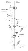
Figure 1
Figure 1
A proposed model for how GPG-NH2 or its metabolites target gp160 for ERAD. Initially, gp160 is co-translationally translocated into the ER, where its growing peptide backbone becomes glycosylated and starts to fold. (1) In the presence of GPG-NH2 or its metabolites gp160 folds incorrectly which targets it to ERAD. (2) Subsequently, gp160 is retro-translocated to the cytoplasm, (3) where it becomes deglycosylated by the cytosolic N-glycanase prior to (4) degradation of its peptide backbone by the proteasome.
Figure 2
Figure 2
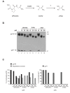
GPG-NH2 and its metabolites G-NH2 and αHGA decrease gp160 mobility and steady-state levels. (A) Scheme of GPG-NH2 being metabolized in cell culture medium supplemented with 10% FBS. GPG-NH2 is processed by CD26 (peptidyl peptidase V) to G-NH2 and subsequently modified into αHGA by an unidentified enzyme. (B) HeLa-tat III cells were transfected to express gp160. Two hours post transfection the cells were treated with the indicated concentrations of GPG-NH2, G-NH2 or αHGA and harvested 20 h post transfection. The cell lysates were separated by SDS-PAGE and immunoblotted with mAb towards gp41. (C) Densitometric measurement of gp160 and degradation products (left panel) and gp41 (right panel) given as percentage of total gp160 or gp41 respectively in untreated cells in (B), lane 1. The results represent the average of two experiments.
Figure 3
Figure 3
αHGA acts on gp160 independently of supplemented serum in cell culture medium. HeLa-tat III cells were cultured in cell culture medium supplemented with 10% FBS and transfected to express gp160 for 20 h. Two hours upon transfection the cell culture supernatants were carefully removed, the cells rinsed twice in PBS and provided with culture medium containing either 10% HS (upper panel) or no serum (lower panel) and indicated concentrations of GPG-NH2, G-NH2 or αHGA. The cell lysates were immunoblotted with mAb towards gp41.
Figure 4
Figure 4
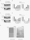
αHGA targets gp160 for degradation more rapidly than GPG-NH2. (A) HeLa-tat III cells were transfected to express gp160 and treated with 20 μM (upper panel) or 100 μM GPG-NH2 (lower panel) for the indicated times pre- or post-transfection. The cells were harvested 24 h post transfection and immunoblotted with mAb towards gp41. (B) Densitometric measurements of gp160 and degradation products in samples treated with 20 μM (left panel) or 100 μM GPG-NH2 (right panel) as described in (A) and given as percentage of total gp160 in untreated cells in (A), lane 1. (C) As in (A), except the cells were treated with αHGA at 20 μM (upper panel) or 100 μM (lower panel). (D) Densitometric measurements as described in (B) of samples treated with αHGA at 20 μM (left panel) or 100 μM (right panel) described in (C). (E) Glycoprotein blot of HeLa-tat III cell lysates collected from cells treated with the indicated concentrations of αHGA for 24 h and stained for total protein and subsequently probed with the lectin Concanavalin A. The asterisks highlight the decreased molecular mass species.
Figure 5
Figure 5
αHGA treatment reduces HIV-1 particle content of Env. (A) Chronically infected ACH-2 cells were induced with PMA for HIV-1 production and treated with the indicated concentrations of αHGA for 72 h. The viral production was determined by measuring extracellular p24 concentrations by ELISA. (B) Virus particles were produced as described in (A) and precipitated with polyethylene glycol followed by immunoblotting towards p24. (C) Immunoblot showing the amount of gp41 present in polyethylene glycol-precipitated HIV-1 particles, produced by ACH-2 as described in (A) for 48 h. The HIV-1 particle content was standardized to the extracellular p24 concentrations measured by ELISA and the gp41/p24 ratio was calculated by densitometry. (D) EM images of immuno-gold labeled gp41 in viral particles surrounding untreated or treated ACH-2 cells with 50 μM αHGA and induced with PMA for 72 h prior to fixation. Arrows indicate labeling of gp41 and the bars represent 100 nm.

Similar articles

References

    1. Land A, Zonneveld D, Braakman I. Folding of HIV-1 envelope glycoprotein involves extensive isomerization of disulfide bonds and conformation-dependent leader peptide cleavage. Faseb J. 2003;17:1058–1067. doi: 10.1096/fj.02-0811com. - DOI - PubMed
    1. Lu M, Blacklow SC, Kim PS. A trimeric structural domain of the HIV-1 transmembrane glycoprotein. Nat Struct Biol. 1995;2:1075–1082. doi: 10.1038/nsb1295-1075. - DOI - PubMed
    1. McCune JM, Rabin LB, Feinberg MB, Lieberman M, Kosek JC, Reyes GR, Weissman IL. Endoproteolytic cleavage of gp160 is required for the activation of human immunodeficiency virus. Cell. 1988;53:55–67. doi: 10.1016/0092-8674(88)90487-4. - DOI - PubMed
    1. Gomez C, Hope TJ. The ins and outs of HIV replication. Cell Microbiol. 2005;7:621–626. doi: 10.1111/j.1462-5822.2005.00516.x. - DOI - PubMed
    1. Kilgore NR, Salzwedel K, Reddick M, Allaway GP, Wild CT. Direct evidence that C-peptide inhibitors of human immunodeficiency virus type 1 entry bind to the gp41 N-helical domain in receptor-activated viral envelope. J Virol. 2003;77:7669–7672. doi: 10.1128/JVI.77.13.7669-7672.2003. - DOI - PMC - PubMed

Publication types