Review
. 2010 Mar 15:5:7.
doi: 10.1186/1749-8104-5-7.

Insulin receptor signaling in the development of neuronal structure and function

Affiliations
Review

Insulin receptor signaling in the development of neuronal structure and function

Shu-Ling Chiu et al. Neural Dev. .

Abstract

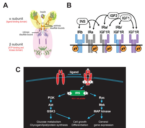
Sensory experience plays a crucial role in regulating neuronal shape and in developing synaptic contacts during brain formation. These features are required for a neuron to receive, integrate, and transmit signals within the neuronal network so that animals can adapt to the constant changing environment. Insulin receptor signaling, which has been extensively studied in peripheral organ systems such as liver, muscle and adipocyte, has recently been shown to play important roles in the central nervous system. Here we review the current understanding of the underlying mechanisms that regulate structural and functional aspects of circuit development, particularly with respect to the role of insulin receptor signaling in synaptic function and the development of dendritic arbor morphology. The potential link between insulin receptor signaling malfunction and neurological disorders will also be discussed.


Figures

Figure 1
Figure 1
Schematic diagram of an excitatory synapse and the temporal sequence of synapse formation and maturation. (A) Synapses are specialized junctions between neurons composed of complex membrane and proteins. A synapse can be divided structurally into three parts: a presynaptic axon terminal packed with synaptic vesicles (SV) and release machinery, a synaptic cleft, and a postsynaptic dendritic counterpart filled with neurotransmitter receptors, scaffold proteins and signaling machinery. (B) Synapse formation is initiated by the contact between dendrites and axons, followed by the recruitment of presynaptic and postsynaptic specializations. Increases in synapse size and synaptic strength by accumulation of AMPA receptors at synapses are characteristics of synapse maturation. AMPAR, α-amino-3-hydroxy-5-methyl-4-isoxazole propionic acid receptor; CaMKII, Calcium calmodulin dependent kinase type II; CASK, calcium calmodulin-dependent serine kinase; GKAP, guanylate kinase-associated protein; GRIP, glutamate receptor-interacting protein; InsP3R, inositol triphosphate receptor; mGluR, metabotropic glutamate receptor; NMDAR, N-methyl-D-aspartate receptor; PSD, postsynaptic density; PSD-95, postsynaptic density protein-95; RIM, Rab3-interacting molecule; SAP, synapse-associated protein; SER, smooth endoplasmic reticulum; SPAR, spine-associated Rap GTPase activating protein; VAMP, vesicle-associated membrane protein; VGCC, voltage-gated calcium channel. (Adapted and modified from [4]).
Figure 2
Figure 2
Dendritic arbor growth and dynamics. (A) Dendrite development can be divided into three phases: phase I, the cell differentiates and extends an axon with little elaboration of the dendritic arbor; phase II, the dendritic arbor grows rapidly; phase III, the dendritic arbor grows slowly and appears stable. Reconstruction of the same tectal neuron imaged in vivo daily for 5 days is shown on top. (Adapted and modified from [19,48].) (B) Dendritic growth is highly dynamic as revealed by time-lapse imaging at 2-hour intervals over a 6-hour imaging period. Stable branches, retracted branches, transient branches, and added branches are shown in gray, red, green and yellow, respectively. Cell bodies are shown in purple. (Adapted from [52].)
Figure 3
Figure 3
Insulin receptor structure and signaling. (A) Insulin receptor monomer, composed of an α (yellow) and β subunit (pink) bridged by an intrinsic disulfide bond, which dimerizes with another insulin receptor monomer through extrinsic disulfide bonds to form a functional receptor. (Adapted and modified from [80].) (B) Ligand selectivity of the insulin receptor homodimer or heterodimer with the insulin-like growth factor (IGF)-1 receptor. Note that the homodimer of the splice variant IRa, the predominant form of inslulin receptor in the brain, binds specifically to insulin (INS), whereas the heterodimer with the IGF-1 receptor binds to not only INS but also IGF-1 and IGF-2. (Adapted and modified from [86].) (C) Insulin receptor signaling initiated by ligand binding activates tyrosine autophosphorylation in the β subunit, which stimulates two major downstream pathways, the phosphoinositide-3 kinase (PI3K)/Akt and Ras/mitogen-activated protein kinase (MAPK) cascades, through insulin receptor substrates (IRSs) and results in a diverse series of cellular processes in peripheral tissues. (Modified from [88].) GSK3, glycogen synthase kinase 3.
Figure 4
Figure 4
Protein sequence alignment of X. laevis insulin receptor IR1 with different species. Amino acid sequence derived from Xenopus IR1 was aligned with rat, mouse and human insulin receptor protein sequences with the ClustalW algorithm. Schematic drawing of the alignment identities (percentages) in different functional domains of insulin receptor. Note that the predicated Xenopus kinase domain shares the highest identity with other species compared to other domains.
Figure 5
Figure 5
Recording of visual responses in an intact animal. (A) X. laevis tadpole. (B) Diagram of the Xenopus visual circuit. Optic tectal neurons receive direct visual input from retinal ganglion cells of the eye. (C) Experimental setup. Visual stimulation was delivered with a green LED pigtailed to an optic fiber that illuminates the entire eye for whole field stimulation. The tadpole brain was cut along the midline to expose the cell bodies. Visual responses from tectal neurons contralateral to the stimulated eye were recorded by whole-cell recording. (D) Visual responses of tectal neurons. Tectal neurons respond to transient light intensity change. The OFF responses induced by light off is normally bigger in amplitude and longer in duration than ON responses induced by light on. Superimposition of 20 consecutive responses (gray) and the averaged trace (black) are shown. Adapted from [84]. dnIR, dominant negative insulin receptor; moIR, morpholino-mediated knockdown of insulin receptor; wtIR, wild-type insulin receptor.
Figure 6
Figure 6
Insulin receptor signaling regulates synapse numbers. (A) Electron micrographs show ultrastructural morphology of synaptic terminals that contact green fluorescent protein (GFP)-, wild-type insulin receptor (wtIR)- and dominant negative insulin receptor (dnIR)-expressing dendrites. The postsynaptic area, presynaptic area and the clustered synaptic vesicle are highlighted in light blue, green, and pink, respectively. (B) dnIR-expressing dendrites receive significantly fewer synaptic contacts compared to GFP- and wtIR-transfected dendrites. (C) Synapses that contact GFP-, wtIR- and dnIR-expressing dendrites show comparable ultrastructural synaptic maturity, determined by the area occupied by clustered synaptic vesicles relative to the area of the presynaptic terminal. (D) Schematic cartoon showing that normal tectal neurons increase total synapse number and, therefore, synaptic transmission, branch stabilization and extension in response to enhanced visual stimulation, whereas tectal neurons expressing dnIR do not increase synapse number and fail to increase synaptic function and dendritic structural plasticity.

Similar articles

Cited by

References

    1. Trachtenberg JT, Chen BE, Knott GW, Feng G, Sanes JR, Welker E, Svoboda K. Long-term in vivo imaging of experience-dependent synaptic plasticity in adult cortex. Nature. 2002;420:788–794. doi: 10.1038/nature01273. - DOI - PubMed
    1. Holtmaat A, Wilbrecht L, Knott GW, Welker E, Svoboda K. Experience-dependent and cell-type-specific spine growth in the neocortex. Nature. 2006;441:979–983. doi: 10.1038/nature04783. - DOI - PubMed
    1. Chen C, Regehr WG. Developmental remodeling of the retinogeniculate synapse. Neuron. 2000;28:955–966. doi: 10.1016/S0896-6273(00)00166-5. - DOI - PubMed
    1. Li Z, Sheng M. Some assembly required: the development of neuronal synapses. Nat Rev Mol Cell Biol. 2003;4:833–841. doi: 10.1038/nrm1242. - DOI - PubMed
    1. Ahmari SE, Buchanan J, Smith SJ. Assembly of presynaptic active zones from cytoplasmic transport packets. Nat Neurosci. 2000;3:445–451. doi: 10.1038/74814. - DOI - PubMed

Publication types

Substances

LinkOut - more resources