Review
. 2010 Mar 16;18(3):342-56.
doi: 10.1016/j.devcel.2010.02.005.

Cellular plasticity within the pancreas--lessons learned from development

Affiliations
Review

Cellular plasticity within the pancreas--lessons learned from development

Sapna Puri et al. Dev Cell. .

Abstract

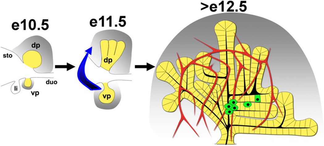
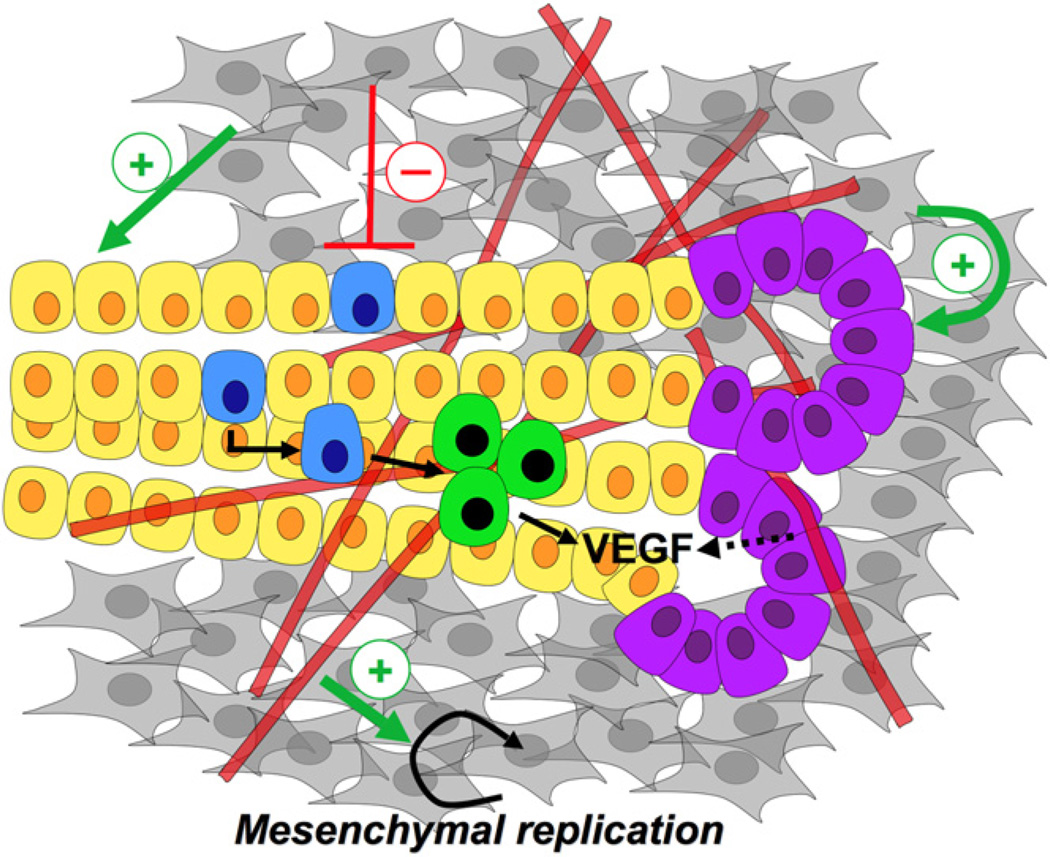
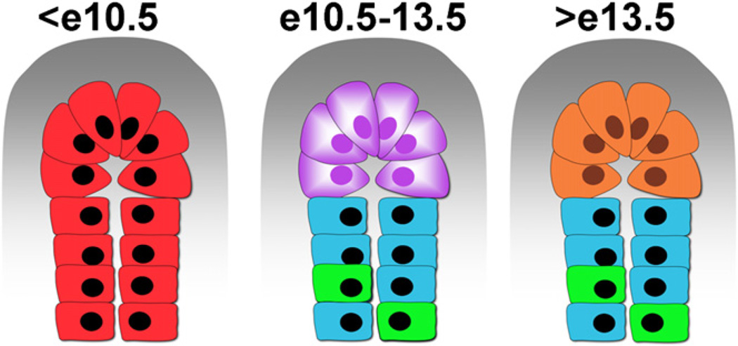
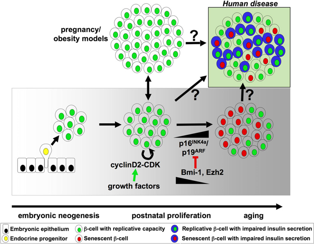
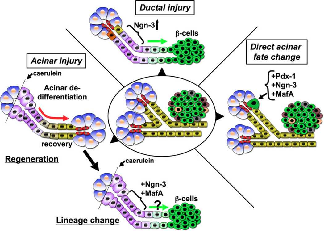
The pancreas has been the subject of intense research due to the debilitating diseases that result from its dysfunction. In this review, we summarize current understanding of the critical tissue interactions and intracellular regulatory events that take place during formation of the pancreas from a small cluster of cells in the foregut domain of the mouse embryo. Importantly, an understanding of principles that govern the development of this organ has equipped us with the means to manipulate both embryonic and differentiated adult cells in the context of regenerative medicine. The emerging area of lineage modulation within the adult pancreas is of particular interest, and this review summarizes recent findings that exemplify how lessons learned from development are being applied to reveal the potential of fully differentiated cells to change fate.


Figures

Figure 1
Figure 1. Early Steps during Embryonic Pancreas Development
Dorsal (dp) and ventral (vp) pancreatic buds can be visualized (yellow) along with the stomach (sto), duodenum (duo), and liver bud (Li) in the posterior foregut at e10.5. The pancreatic mesenchyme (gray) collects around developing buds and provides essential instructive signals. Gut rotation (represented by the blue arrow) at e11.5 leads to fusion of the ventral and dorsal aspects followed by expansion into the surrounding mesenchyme (gray). After e12.5, secondary transition leads to endocrine specification (green) within the epithelium furthest from the mesenchyme, in close apposition to the vasculature (red vessels).
Figure 2
Figure 2. The Developmental Niche of the Embryonic Pancreas
During the early stages of development (e10.5–e13.5), multipotent progenitor cells within the pancreatic epithelium (yellow) expand into the surrounding mesenchyme (gray) while being populated by the endothelium (red tubes). Epithelium-mesenchyme, epithelium-endothelium, and endothelium-mesenchyme interactions occur simultaneously during pancreas development. The mesenchyme positively promotes the ‘‘proximal’’ tissue (purple cells), while being inhibitory to the more ‘‘distal,’’ endocrine progenitor cells (blue), which have been observed to form closer to the interior of the three-dimensional organ. Interestingly, the mesenchyme has a positive influence on the pancreatic progenitors. Endocrine cells (green) are found in close apposition to the vasculature, and known to secrete VEGF, which may recruit more blood vessels to the site of islet formation. The endothelium also exerts a positive effect on the mesenchyme, as in the absence of the vasculature the mesenchyme fails to develop around the dorsal pancreatic bud.
Figure 3
Figure 3. Lineage Restriction within the Pancreatic Epithelium
Expansion ofthe progenitorpool atthe tip of the epithelium occurs early during development. From an uncommitted pool of progenitors (before e10.5, in red) to multipotent, Cpa-1-positive cells (purple) that give rise to exocrine (orange), ductal (blue), and endocrine cells (green). Endocrine progenitors go on to delaminate from the epithelium and cluster with other endocrine cells. It is believed that distance from the mesenchyme influences the decision of exocrine versus endocrine lineage.
Figure 4
Figure 4. β-Cell Expansion
During embryonic pancreas development, β-cell (green) expansion occurs through neogenesis from progenitors (yellow) within the pancreatic epithelium (black). In postnatal life, under normal conditions, β-cell proliferation through the cyclinD2-CDK complex is the predominant mode to adjust for changes in insulin demand, e.g., during pregnancy or development of obesity. With age, however, accumulation of tumor suppressors p16INK4a/p19ARF leads to a block in the replication potential of β-cells (red) concomitant with a reduction in the epigenome modifiers Bmi-1 and Ezh2. Although it is unclear what the role of β-cell replication and senescence is in the development ofhuman disease suchas type 2diabetes, one can envision an islet populated by cells competent to divide and respond to glucose and those lacking these abilities (Weir et al., 2009). Other endocrine cells are not shown for simplicity.
Figure 5
Figure 5. Fate Manipulation within the Adult Pancreas
In a ductal injury model such as pancreatic ductal ligation (top schematic), normal duct cells (yellow), or duct-associated cells (orange) adopt a progenitor state (purple) and upregulate Ngn-3 expression that correlates with increased β-cell formation, depicted by the green arrow. In an acinar cell injury model (left schematic), acinar cells (blue) undergo dedifferentiation (purple) (for e.g., upon caerulein treatment) followed by recovery (red arrow) and thus regeneration. Introduction of key transcription factors such as Ngn-3 and MafA into such dedifferentiated cells, shown in the bottom schematic, might promote β-cell fate. A third scenario is of direct acinar fate change (right panel). Ectopic expression of Pdx-1, Ngn-3, and MafA in the adult pancreas redirects acinar cells to adopt an identity resembling an endocrine β-cell (green), although these cells fail to cluster with other endocrine cells.

Similar articles

Cited by

References

    1. Ahlgren U, Jonsson J, Edlund H. The morphogenesis of the pancreatic mesenchyme is uncoupled from that of the pancreatic epithelium in IPF1/PDX1-deficient mice. Development. 1996;122:1409–1416. - PubMed
    1. Apelqvist A, Ahlgren U, Edlund H. Sonic hedgehog directs specialised mesoderm differentiation in the intestine and pancreas. Curr. Biol. 1997;7:801–804. - PubMed
    1. Apelqvist A, Li H, Sommer L, Beatus P, Anderson DJ, Honjo T, Hrabe de Angelis M, Lendahl U, Edlund H. Notch signalling controls pancreatic cell differentiation. Nature. 1999;400:877–881. - PubMed
    1. Attali M, Stetsyuk V, Basmaciogullari A, Aiello V, Zanta-Boussif MA, Duvillie B, Scharfmann R. Control of beta-cell differentiation by the pancreatic mesenchyme. Diabetes. 2007;56:1248–1258. - PubMed
    1. Baeyens L, Bonne S, Bos T, Rooman I, Peleman C, Lahoutte T, German M, Heimberg H, Bouwens L. Notch signaling as gatekeeper of rat acinar-to-beta-cell conversion in vitro. Gastroenterology. 2009;136:1750–1760. - PubMed

Publication types

LinkOut - more resources