. 2010 Mar 16;18(3):385-96.
doi: 10.1016/j.devcel.2009.12.024.

Hsk1- and SCF(Pof3)-dependent proteolysis of S. pombe Ams2 ensures histone homeostasis and centromere function

Affiliations

Hsk1- and SCF(Pof3)-dependent proteolysis of S. pombe Ams2 ensures histone homeostasis and centromere function

Yuko Takayama et al. Dev Cell. .

Abstract

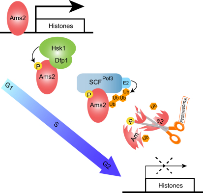
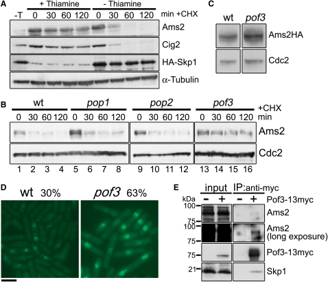
Schizosaccharomyces pombe GATA factor Ams2 is responsible for cell cycle-dependent transcriptional activation of all the core histone genes peaking at G1/S phase. Intriguingly, its own protein level also fluctuates concurrently. Here, we show that Ams2 is ubiquitylated and degraded through the SCF (Skp1-Cdc53/Cullin-1-F-box) ubiquitin ligase, in which F box protein Pof3 binds this protein. Ams2 is phosphorylated at multiple sites, which is required for SCF(Pof3)-dependent proteolysis. Hsk1/Cdc7 kinase physically associates with and phosphorylates Ams2. Even mild overexpression of Ams2 induces constitutive histone expression and chromosome instability, and its toxicity is exaggerated when Hsk1 function is compromised. This is partly attributable to abnormal incorporation of canonical H3 into the central CENP-A/Cnp1-rich centromere, thereby reversing specific chromatin structures to apparently normal nucleosomes. We propose that Hsk1 plays a vital role during post S phase in genome stability via SCF(Pof3)-mediated degradation of Ams2, thereby maintaining centromere integrity.


Figures

None
Graphical abstract
Figure 1
Figure 1
Ams2 Protein Is Stable during S Phase (A) Stability of the Ams2 protein at various stages of the cell cycle. The experimental scheme is shown in the upper panel. Wild-type, cdc10-129, and cdc2-33 cells carrying the plasmid pREP41-Ams2 were treated with thiamine and CHX (time 0). Protein samples were prepared at the indicated time points. Immunoblotting was performed with Ams2 (upper) and TAT-1 (lower) antibodies. See also Figure S1. (B–D) Quantification of Ams2 protein stability. Band intensities of Ams2 shown in (A) were quantified by normalization relative to α-tubulin signals. Relative intensity at time 0 was set as 100% in each panel. (E) Ams2 stability during M phase. The experimental scheme is shown in the top panel. Relative intensities are shown for wild-type (open circles) and nda3-KM311 (closed circles) cells.
Figure 2
Figure 2
Ams2 Is Polyubiquitylated and Degraded by the Proteasome Pathway (A) Stabilization of Ams2 in the proteasome mutant. Wild-type and mts2-1 cells carrying pREP41-Ams2 were grown and cell extracts were prepared as in Figure 1. The data for wild-type were adopted from Figure 1A. See also Figure S1. (B) Quantification of Ams2 protein stability. Band intensities of Ams2 were measured and normalized as in Figure 1. (C) Ubiquitylation of Ams2. Episomal His6-ubiquitin (His-Ub, lanes 1 and 3) and HA-ubiquitin (HA-Ub, lanes 2 and 4) were expressed in wild-type (lanes 1 and 2) and mts2-1 (lanes 3 and 4) cells. Ubiquitylated proteins were purified, followed by immunoblotting with Ams2 antibody. See also Figure S2.
Figure 3
Figure 3
Ams2 Is Degraded via SCFPof3 Ubiquitin Ligase (A) Stabilization of Ams2 in a Skp1 shut-off strain. Cells containing thiamine-repressible nmtP3-HA-skp1+ were grown in the absence of thiamine and cultures were divided into two parts at time 0. CHX was added to both cultures, while thiamine was added to one part (± Thiamine). Immunoblotting was then performed with Ams2, Cig2, HA, and α-tubulin antibodies. (B) Stabilization of Ams2 in pof3 mutants. Wild-type, pop1, pop2, or pof3 cells were treated with CHX (time 0). Ams2 stability was analyzed by immunoblotting with anti-Ams2 (upper). Cdc2 levels were used as a loading control. See also Figure S3. (C) Elevated levels of Ams2 in pof3 mutants. Protein extracts were prepared from wild-type and pof3 cells whose ams2+ gene was tagged by HA epitope. Ams2 protein levels were measured by immunoblotting with HA antibody (upper). Cdc2 levels were used as a loading control (lower). (D) Accumulation of Ams2-GFP in the absence of Pof3. Wild-type (left) and pof3 cells (right) containing Ams2-GFP expressed under the endogenous promoter were fixed in formaldehyde, and GFP signals were examined under fluorescence microscopy. Exposure time was the same for both strains. The percentages of uninucleate cells showing nuclear Ams2-GFP signals were counted (n > 200 cells). Note that pof3 mutant cells were longer than wild-type controls because of DNA damage checkpoint-dependent G2 delay (Katayama et al., 2002). Scale bar represents 10 μm. (E) Physical interaction between Ams2 and Pof3. mts3-1 pof3+-13myc (+) or mts3-1 (−) cell lysates were immunoprecipitated with anti-myc antibody-coupled affinity resin. Precipitated proteins were detected by immunoblotting with myc, Ams2, and Skp1 antibodies.
Figure 4
Figure 4
Phosphorylation of Ams2 Renders the Protein Unstable (A) Phosphorylation of Ams2. Cell extracts prepared from untagged wild-type (no-tag) or HA-tagged (Ams2-HA) cells were immunoprecipitated with anti-HA, followed by incubation with λ-PPase (lanes 2 and 4) or heat-denatured λ-PPase (lane 5). (B) Schematic representation of phosphorylation motifs in Ams2. Vertical black lines show zinc finger motifs. Amino acid sequences are shown with the corresponding sequences of Ams2 homologs from different Schizosaccharomyces species. S.p., S. pombe; S.j., S. japonicus; S.o., S. octosporus. Identical amino acids (gray shaded), phosphorylation motifs (asterisks), and Cdc2 phosphorylation consensus motifs (boxes) are shown. See also Figure S4. (C) Stabilization of alanine-substituted Ams2 proteins. Wild-type cells carrying pREP41-Ams2 or Ams2 alanine mutants were grown in the absence of thiamine. Experiments were performed in a manner similar to that described in Figure 1 except that cultures were incubated at 33°C. (D) Reduced phosphorylation of alanine-substituted Ams2 mutants. Cells containing alanine-substituted Ams2-HA expressed under the endogenous promoter were arrested at S phase by HU. Cell extracts were immunoprecipitated with HA antibody and treated with λ-PPase (lanes 2, 5, 8, 11, and 14) or heat-denatured λ-PPase (lanes 3, 6, 9, 12, and 15).
Figure 5
Figure 5
Hsk1 Is a Major Kinase Required for Ams2 Phosphorylation (A and B) Reduced phosphorylation of Ams2 in the hsk1 mutant. (A) Exponentially growing cultures of the indicated strains were incubated for 5 hr at 25°C or 37°C in the presence of HU. Immunoblotting was performed with Ams2 (upper) or TAT-1 (lower) antibodies. See also Figure S5A. (B) Cell extracts prepared from wild-type or hsk1-89 were immunoprecipitated with HA antibody and treated with λ-PPase (lanes 2 and 5) or heat-denatured λ-PPase (lanes 3 and 6). Immunoblotting was performed with Ams2 antibody. (C) Hypophosphorylated Ams2 is stabilized in the hsk1 mutant. Wild-type and hsk1-89 cells were incubated for 3 hr at 37°C, followed by CHX treatment. Slow migrating forms evident only in wild-type cells are indicated by arrows. (D) Ams2 interacts with Dfp1. The indicated plasmids were introduced into L40 and three independent transformants were assayed for β-galactosidase activity. See also Figure S5B. (E) Ams2 coimmunoprecipitates with Hsk1-Dfp1. Protein extract was prepared from cells expressing FLAG-tagged Hsk1 and HA-tagged Ams2 and immunoprecipitation was performed with FLAG or HA antibodies after formaldehyde crosslinking. Precipitated proteins were detected by immunoblotting with individual antibodies. (F) Ams2 is phosphorylated by DDK. In vitro kinase assay was performed and reaction proteins were separated on SDS-PAGE and detected by autoradiography. As substrates, His-tagged fusion proteins were visualized by Coomassie blue staining (CBB). The arrow indicates the position of the full-length Ams2 fusion proteins.
Figure 6
Figure 6
Ams2 Overproduction Leads to Constitutive Expression of Histone Genes in Wild-Type Cells and Is Deleterious in the hsk1-89 Mutant (A) Ams2 overproduction results in cell toxicity. Wild-type cells containing pREP1, pREP41, pREP1-Ams2, or pREP41-Ams2 were grown on minimal medium plates in the absence of thiamine for 3 days at 33°C. The hsk1-89 mutant cells containing pREP41 or pREP41-Ams2 were grown on minimal medium plates in the absence of thiamine for 4 days at 26°C. See also Figure S6A. (B) Growth curve of Ams2 overproduced cells. Wild-type cells carrying pREP1 or pREP1-Ams2 were grown in minimal medium in the presence (OFF) or absence (ON) of thiamine at 33°C. At the indicated times, aliquots were withdrawn to count cell numbers. (C) Overproduced Ams2 leads to cell cycle-independent histone gene expression. Wild-type (WT), ams2, cdc10-129, and cdc2-33 cells carrying pREP41-Ams2 plasmids grown in the absence (Ams2 ON) or presence (Ams2 OFF) of thiamine at 26°C were shifted to 36°C for 3 hr or HU was added concomitant with temperature shift-up. The expression levels of histone H2A+, H2B+, H3+, H4+, act1+, and ams2+ genes were examined by northern blotting. See also Figure S6B. (D) Levels of histone mRNAs increase in asynchronous cells expressing alanine-substituted Ams2 mutant proteins and hsk1 mutant cells. The expression levels of histone H2A+, H2B+, H3+, H4+, and act1+ genes were examined by northern blotting. Total RNA samples were prepared from wild-type cells carrying pREP41, pREP41-Ams2, or pREP41-Ams2 M2&M3 and grown in the absence of thiamine (left panel). In the right panel, samples were prepared from wild-type or hsk1-89 cells incubated for 4 hr at 26°C, 30°C, or 37°C. (E) Levels of histone and ams2+ mRNAs in asynchronous cells expressing alanine-substituted (M2&M3) Ams2 mutant proteins from its native promoter. Total RNA samples were prepared from cells whose genomic ams2+ ORF was replaced with either ams2WT-HA or ams2M2&M3-HA, and the expression levels of histones, act1+, and ams2+ genes were examined by northern blotting. The amount of ams2+ mRNA were quantified with a phosphorImager (Typhoon 9410 imager, GE Healthcare) and normalized against that of act1+ mRNA. (F) Minichromosome loss assay. Wild-type or Ams2-induced (pnmt81-Ams2) cells carrying Ch10-CN2 minichromosome were spread on minimal medium plates containing appropriate supplements (low concentration of adenine) without thiamine and incubated for 4 days at 33°C. Data are shown as means ± SD from six independent experiments.
Figure 7
Figure 7
Overproduction of Ams2 Disrupts Central Centromere Structures (A) The overall centromere structure (chromosome I) is depicted with the cnt probe used (bar). Chromatin fractions were prepared from wild-type cells carrying pREP1 or pREP1-Ams2 in the presence (OFF) or absence (ON) of thiamine (18 hr induction, 4 gels on the left; 20 hr induction, gels on the right). These samples were treated with MNase for 0, 1, 2, 5, 10 (4 gels on the left), and 20 min (right), followed by agarose gel electrophoresis (EtBr) and Southern blotting with the central centromere probe (cnt). (B) Abnormal H3 localization at the central centromere cells. Cell extracts were prepared from wild-type cells carrying pREP1 or pREP1-Ams2 in the presence (OFF) or absence (ON) of thiamine (18 hr induction). DNAs coimmunoprecipitated with cnp1, H3, or H4 antibodies were quantified by real-time PCR with act1 and cnt2 probes. The amount of each immunoprecipitated DNA was divided by that of the corresponding whole-cell extract DNA after background titration. See also Figure S7. (C) Model explaining how histone levels and central centromere structures are maintained via Hsk1- and SCFPof3-mediated degradation of Ams2. In wild-type cells (top), Ams2 accumulates only during G1 and S, which maintains both cell cycle-dependent histone stoichiometry and a proper balance between Cnp1/H4 (C, orange circles) and H3/H4 (3, blue circles). On the other hand, when Ams2 is overproduced (e.g., hsk1-89), excess Ams2 induces cell cycle-independent synthesis of histones, leading to centromere malfunction and chromosome instability.

Comment in

Similar articles

Cited by

References

    1. Anderson H.E., Wardle J., Korkut S.V., Murton H.E., Lopez-Maury L., Bähler J., Whitehall S.K. The fission yeast HIRA histone chaperone is required for promoter silencing and the suppression of cryptic antisense transcripts. Mol. Cell. Biol. 2009;29:5158–5167. - PMC - PubMed
    1. Baber-Furnari B.A., Rhind N., Boddy M.N., Shanahan P., Lopez-Girona A., Russell P. Regulation of mitotic inhibitor Mik1 helps to enforce the DNA damage checkpoint. Mol. Biol. Cell. 2000;11:1–11. - PMC - PubMed
    1. Bailis J.M., Bernard P., Antonelli R., Allshire R.C., Forsburg S.L. Hsk1-Dfp1 is required for heterochromatin-mediated cohesion at centromeres. Nat. Cell Biol. 2003;5:1111–1116. - PubMed
    1. Blackwell C., Martin K.A., Greenall A., Pidoux A., Allshire R.C., Whitehall S.K. The Schizosaccharomyces pombe HIRA-like protein Hip1 is required for the periodic expression of histone genes and contributes to the function of complex centromeres. Mol. Cell. Biol. 2004;24:4309–4320. - PMC - PubMed
    1. Brown G.W., Kelly T.J. Purification of Hsk1, a minichromosome maintenance protein kinase from fission yeast. J. Biol. Chem. 1998;273:22083–22090. - PubMed

Publication types

MeSH terms