. 2010 Mar 16;18(3):410-24.
doi: 10.1016/j.devcel.2009.12.022.

Ofd1, a human disease gene, regulates the length and distal structure of centrioles

Affiliations

Ofd1, a human disease gene, regulates the length and distal structure of centrioles

Veena Singla et al. Dev Cell. .

Abstract

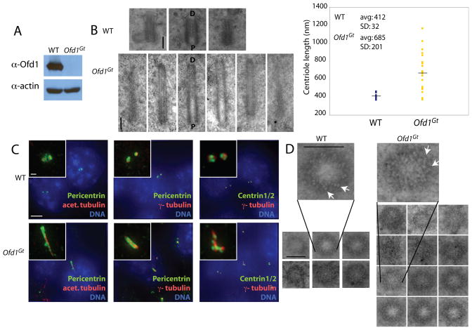
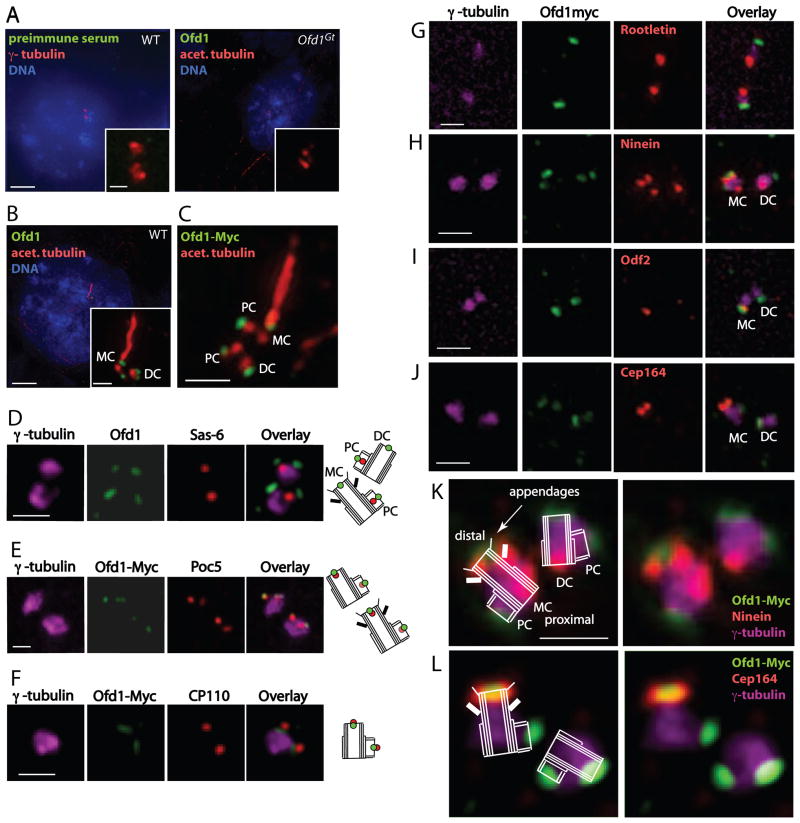
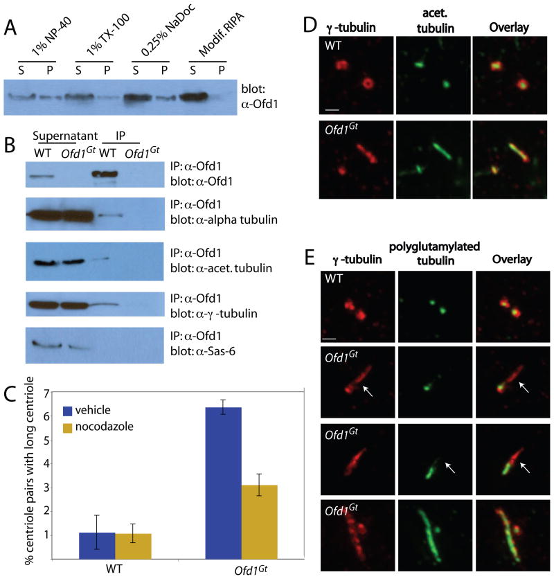
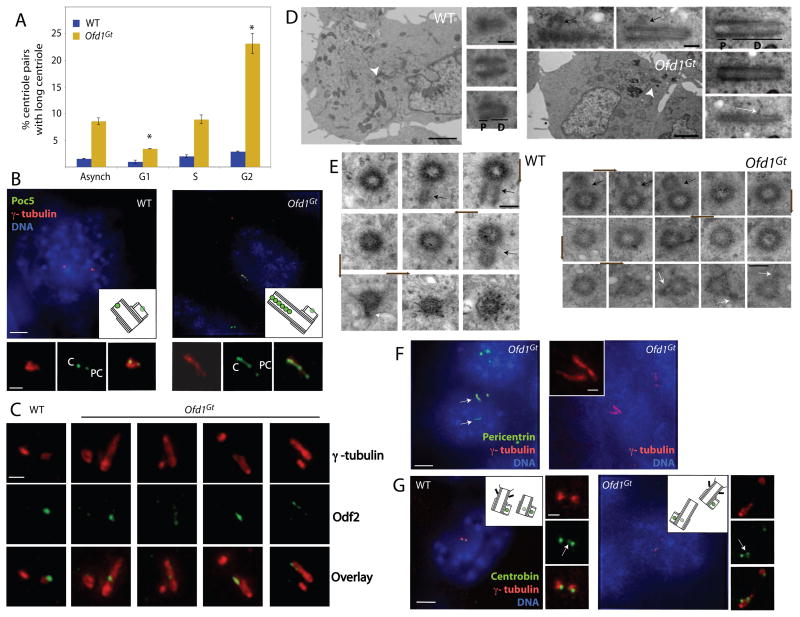
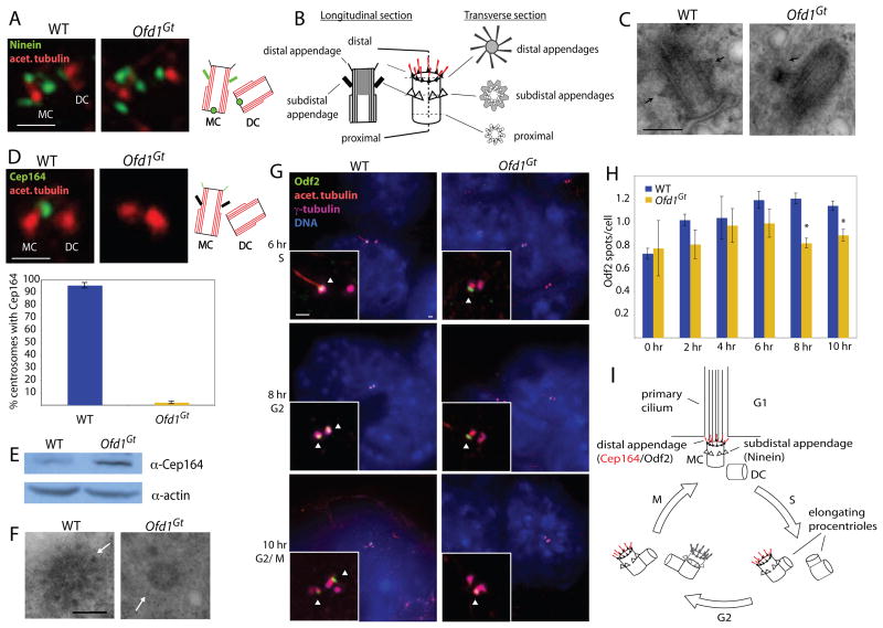
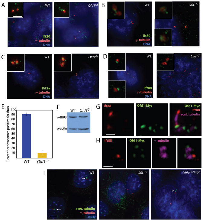
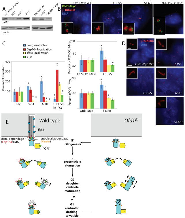
Centrosomes and their component centrioles represent the principal microtubule organizing centers of animal cells. Here, we show that the gene underlying orofaciodigital syndrome 1, Ofd1, is a component of the distal centriole that controls centriole length. In the absence of Ofd1, distal regions of centrioles, but not procentrioles, elongate abnormally. These long centrioles are structurally similar to normal centrioles but contain destabilized microtubules with abnormal posttranslational modifications. Ofd1 is also important for centriole distal appendage formation and centriolar recruitment of the intraflagellar transport protein Ift88. To model OFD1 syndrome in embryonic stem cells, we replaced the Ofd1 gene with missense alleles from human OFD1 patients. Distinct disease-associated mutations cause different degrees of excessive or decreased centriole elongation, all of which are associated with diminished ciliogenesis. Our results indicate that Ofd1 acts at the distal centriole to build distal appendages, recruit Ift88, and stabilize centriolar microtubules at a defined length.


Figures

Figure 1
Figure 1. Ofd1 is essential for centriole length control
(A) Immunoblot of cell lysate supernatants from wild type (WT) and Ofd1Gt cells. 15 μg protein loaded per lane. (B) Longitudinal TEM sections of WT and Ofd1Gt cell centrioles. Long centrioles (defined as > 600 nm) are seen in 35% of Ofd1Gt cells. P, proximal end and D, distal end of centriole. Graph shows centriole length data, collected from 9 WT and 23 Ofd1Gt centrioles. Each measured centriole was from a distinct cell. (C) Representative fluorescence micrographs of WT and Ofd1Gt cells showing centrosomes (Pericentrin and γ-tubulin), centrioles (Centrin and acetylated tubulin), and DNA (DAPI). (D) Transverse TEM sections of WT and Ofd1Gt cell centrioles. White arrows indicate triplet microtubules. Normal length centrioles are contained within a maximum of 8–10 transverse sections, whereas long centrioles span more than 10 sections. Scale bars indicate 200 nm (TEM), 5 μm, and 1 μm (inset). See also Figure S1.
Figure 2
Figure 2. Ofd1 localizes to the distal ends of mother, daughter, and procentrioles
(A–C) Representative micrographs of WT and Ofd1Gt cells showing centrosomes (γ-tubulin), centrioles and cilia (acetylated tubulin), DNA (DAPI), and other indicated antibodies. (D) WT cells showing centrosomes (γ-tubulin), Ofd1, and the proximal procentriole (Sas-6). (E) Ofd1Ofd1myc cells showing centrosomes (γ-tubulin), Myc (Ofd1-Myc), and the distal procentriole (Poc5). Poc5 localizes more strongly to mother or daughter centrioles than to procentrioles. (F) Ofd1Ofd1myc cells showing centrosomes (γ-tubulin), Myc (Ofd1-Myc), and the distal centriole and procentriole (CP110). (G) Ofd1Ofd1myc cells showing centrosomes (γ-tubulin), Myc (Ofd1-Myc), and the proximal centriole (Rootletin). (H) Ofd1Ofd1myc cells showing centrosomes (γ-tubulin), Myc (Ofd1-Myc), and mother centriole subdistal appendages (Ninein). The mother centriole is marked by 3 Ninein foci (2 on the subdistal appendages and one on the proximal end) whereas the daughter centriole is marked by one Ninein focus (on the proximal end). (I) Ofd1Ofd1myc cells showing centrosomes (γ-tubulin), Myc (Ofd1-Myc), and mother centriole appendages (Odf2). (J) Ofd1Ofd1myc cells showing Myc (Ofd1-Myc), mother centriole distal appendages (Cep164), and centrosomes (γ-tubulin). (K) – (L) Schematics showing Ofd1Ofd1myc cells stained for Myc (Ofd1-Myc), mother centriole subdistal (Ninein) or distal (Cep164) appendages, and centrosomes (γ-tubulin). MC, mother centriole. DC, daughter centriole. PC, procentriole. Scale bars (A)–(B) indicate 5 μm and 1 μm (inset), and 1 μm for (C)–(L). See also Figure S2.
Figure 3
Figure 3. Ofd1 complexes contain centriolar microtubule components and control centriole microtubule stability
(A) Immunoblot showing Ofd1 (detected with an Ofd1 antibody) in the supernatant (S) and pellet (P) of WT cells lysed with various detergents. (B) Immunoblots of Ofd1 complexes immunoprecipitated from WT or Ofd1Gt cell supernatant with an Ofd1 antibody. (C) Graph indicating percent of long centrioles in WT and Ofd1Gt cells. Cells were treated with nocodazole, fixed and stained for α- and γ-tubulin. γ-tubulin foci more than twice as long as they were wide were counted as long centrioles. Because immunofluorescent (IF) microscopy has lower resolution than TEM, a smaller percent of Ofd1Gt centrioles appeared long when assessed by IF (6–10% by IF versus 35% by TEM). (D) WT and Ofd1Gt cells stained for centrosomes (γ-tubulin) and acetylated tubulin. (E) WT and Ofd1Gt cells stained for centrosomes (γ-tubulin) and polyglutamylated tubulin (GT335). Arrows indicate areas of reduced or absent polyglutamylation. Scale bars indicate 1μm.
Figure 4
Figure 4. Ofd1 restrains growth of the distal domain of both mother and daughter centrioles in G2
(A) Graph indicating the percent of long centrioles in WT and Ofd1Gt cells in different cell cycle phases. Asterisks indicate statistically significant differences compared to the asynchronous population (p < 0.01). (B) The distal centriole (Poc5), centrosomes (γ-tubulin), and DNA (DAPI) of WT and Ofd1Gt cells. Poc5 localizes more strongly to mother or daughter centrioles than to procentrioles. PC, procentriole. C, centriole. (C) Centriole appendages (Odf2) and centrosomes (γ-tubulin) of WT and Ofd1Gt cells. (D) Longitudinal and (E) transverse TEM sections of a WT and long Ofd1Gt centriole. Centriole proximal domain (P), distal domain (D), subdistal appendages (white arrows), procentrioles (black arrows). Arrowheads indicate centrioles in low magnification TEM images. Brown arrows show direction of section sequence. Normal length centrioles are contained within 8–10 sequential transverse sections, whereas long centrioles span more than 10 sections. (F) Ofd1Gt cells showing centrosomes (Pericentrin and γ-tubulin) and DNA (DAPI). (G) Daughter centrioles and procentrioles (Centrobin), centrosomes (γ-tubulin), and DNA (DAPI) of WT and Ofd1Gt cells. In S-G2 phase, Centrobin localizes more strongly to the procentrioles than to the daughter centriole. Arrows indicate daughter centrioles. Scale bars indicate 2 μm (TEM, low magnification), 200 nm (TEM, high magnification), 5 μm and 1 μm (inset). See also Figure S3.
Figure 5
Figure 5. Ofd1 is essential for distal appendage formation
(A) Centrioles (acetylated tubulin) and mother centriole subdistal appendages (Ninein) of WT and Ofd1Gt cells. (B) Diagram depicting the TEM appearance of a mother centriole in transverse and longitudinal views. (C) TEM longitudinal views of WT and Ofd1Gt centrioles. Arrows indicate subdistal appendages. (D) Centrioles (acetylated tubulin) and mother centriole distal appendages (Cep164) of WT and Ofd1Gt cells. Graph shows percent of centrosome pairs showing Cep164 localization in WT and Ofd1Gt cells. (E) Immunblot showing Cep164 in the supernatants of WT and Ofd1Gt cell lysates. 20 μg protein loaded per lane. (F) TEM transverse views of WT and Ofd1Gt centrioles. Arrows indicate distal appendages. (Full serial reconstructions are included in Figure S4). (G) Centrioles and cilia (acetylated tubulin), centriole appendages (Odf2), centrosomes (γ-tubulin), and DNA (DAPI) of WT and Ofd1Gt cells at the indicated time after release from cell synchronization block. Arrowheads indicate centrioles positive for Odf2. (H) Quantification of Odf2 foci per cell in WT and Ofd1Gt cells at the indicated time after release from cell synchronization block. Asterisks indicate p< 0.05. (I) Diagram showing centriole duplication and maturation in coordination with the cell cycle. MC, mother centriole. DC, daughter centriole. Scale bar indicates 200 nm (TEM) and 1 μm. See also Figure S4.
Figure 6
Figure 6. Ofd1 is required for centrosomal recruitment of Ift88, but not Ift20, Ift80, or Kif3a
(A) The intraflagellar transport protein Ift20, centrosomes (γ– tubulin), and DNA (DAPI) of WT and Ofd1Gt cells. IFT20 localizes to the Golgi and near the centrosome (Follit et al., 2006). (B) Ift80, centrosomes (γ–tubulin), and DNA (DAPI) of WT and Ofd1Gt cells. (C) Anterograde kinesin motor component Kif3A, centrosomes (γ–tubulin), and DNA (DAPI) of WT and Ofd1Gt cells. (D) Ift88, centrosomes (γ–tubulin), and DNA (DAPI) of WT and Ofd1Gt cells. (E) Graph showing percent of centrosome pairs with Ift88 localization in WT and Ofd1Gt cells. (F) Immunblot showing Ift88 in the supernatants of WT and Ofd1Gt cell lysates. 20 μg protein loaded per lane. (G) Ift88, Ofd1-Myc (Myc), and centrioles and cilia (acetylated tubulin) of Ofd1Ofd1myc cells. (H) Ift88, Ofd1-Myc (Myc), and centrosomes (γ-tubulin) of Ofd1Ofd1myc cells. (I) Centrioles and cilia (acetylated tubulin), centrosomes (γ-tubulin), and DNA (DAPI) of WT, Ofd1Gt and Ofd1Ofd1myc cells. Arrows indicate cilia. Scale bar indicates 5 μm or 1 μm (inset, (G)- (H)). See also Figure S5.
Figure 7
Figure 7. Missense Ofd1 mutations found in human patients affect centriole length control and ciliogenesis
(A) Immunoblots showing Ofd1 (detected with an Ofd1 antibody) in the supernatants of lysates from cells of the indicated genotypes. 20 μg protein loaded per lane. (B) Cells of the indicated genotypes stained for Ofd1-Myc (Myc), centrosomes (γ-tubulin), and DNA (DAPI). (C) Graphs comparing the frequencies of long centriole formation, centriolar localization of Cep164 and Ift88, and ciliogenesis of cells with Ofd1 alleles to Ofd1Rev, Ofd1IRESOfd1myc, or Ofd1Ofd1myc cells. Asterisks indicate statistically significant differences (p < 0.05). (D) Cells of the indicated genotypes showing centrosomes (γ-tubulin) and DNA (DAPI). (E) A model of Ofd1-dependent control of centriole structure. In wild type cells, the mother centriole distal appendages contain Odf2 and Cep164, and Ift88 is recruited at the distal centriole for entry into the primary cilium. Ofd1 binds to the distal ends of centriolar microtubules, stabilizes centrioles at the proper length during maturation and recruits Ift88 and distal appendage proteins. In the absence of Ofd1, both mother and daughter centrioles show microtubule destabilization and unrestrained elongation of the distal domain during G2, the phase during which centriole maturation occurs. Without Ofd1, subdistal and distal appendages may be present in the middle of the long centriole, or distributed along the elongated portion. The inability to make primary cilia may be due to centriole elongation defects, distal appendage defects, Ift88 recruitment defects, inability to dock to a vesicular membrane (Sorokin, 1962), or a combination of these. See also Figure S7.

References

    1. Azimzadeh J, Bornens M. Structure and duplication of the centrosome. J Cell Sci. 2007;120:2139–2142. - PubMed
    1. Azimzadeh J, Hergert P, Delouvee A, Euteneuer U, Formstecher E, Khodjakov A, Bornens M. hPOC5 is a centrin-binding protein required for assembly of full-length centrioles. J Cell Biol. 2009;185:101–114. - PMC - PubMed
    1. Badano JL, Mitsuma N, Beales PL, Katsanis N. The ciliopathies: an emerging class of human genetic disorders. Annu Rev Genomics Hum Genet. 2006;7:125–148. - PubMed
    1. Badano JL, Teslovich TM, Katsanis N. The centrosome in human genetic disease. Nat Rev Genet. 2005;6:194–205. - PubMed
    1. Bahe S, Stierhof YD, Wilkinson CJ, Leiss F, Nigg EA. Rootletin forms centriole-associated filaments and functions in centrosome cohesion. J Cell Biol. 2005;171:27–33. - PMC - PubMed

Publication types

MeSH terms