. 2010 Mar 16;18(3):425-36.
doi: 10.1016/j.devcel.2010.01.015.

Sorting of the Alzheimer's disease amyloid precursor protein mediated by the AP-4 complex

Affiliations

Sorting of the Alzheimer's disease amyloid precursor protein mediated by the AP-4 complex

Patricia V Burgos et al. Dev Cell. .

Abstract

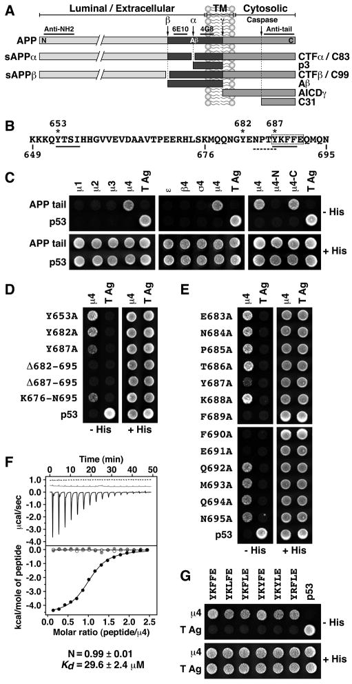
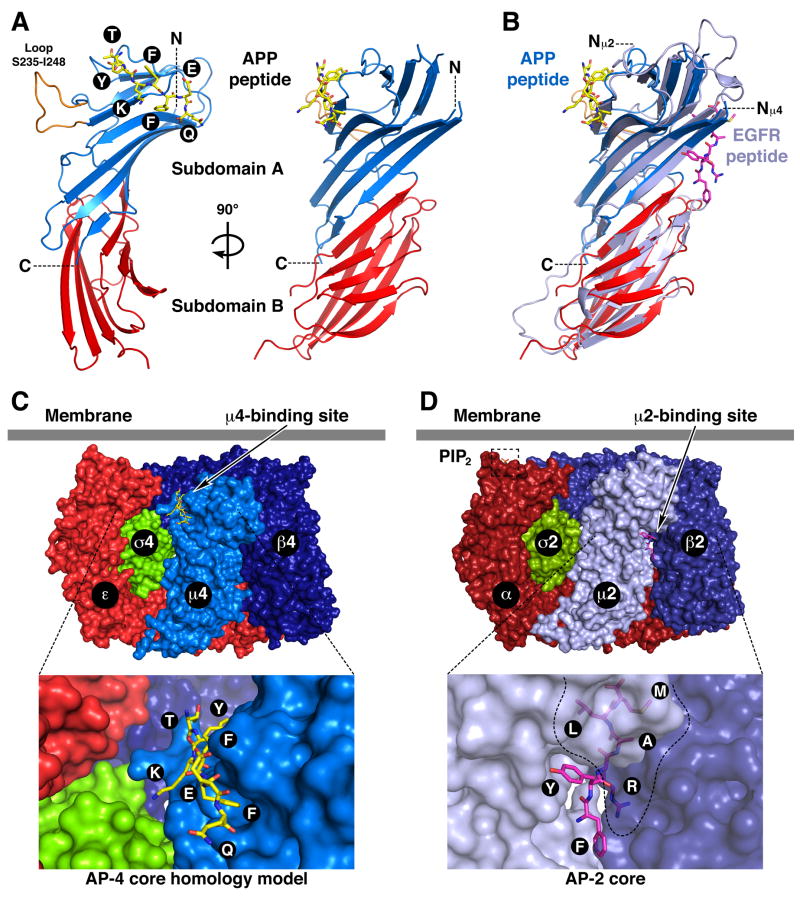
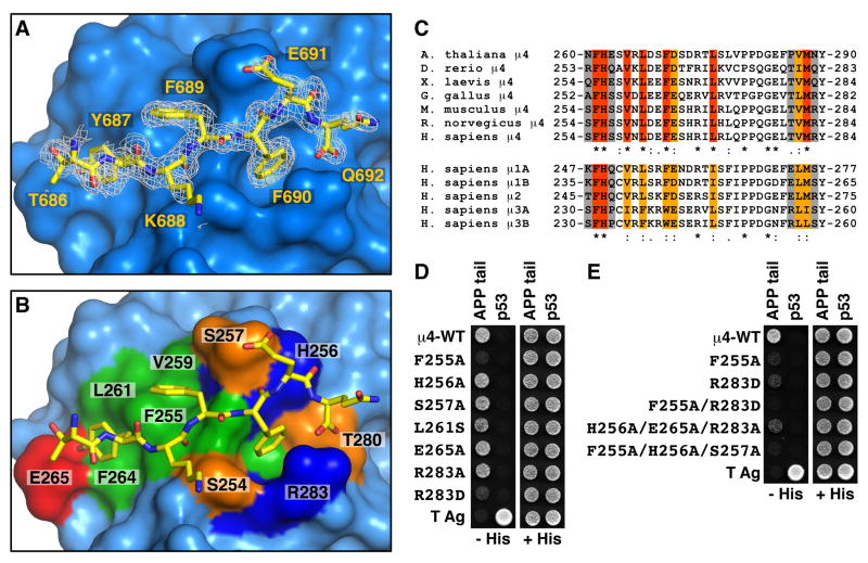
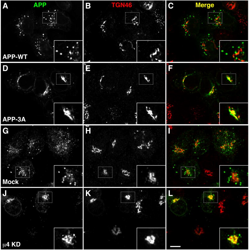
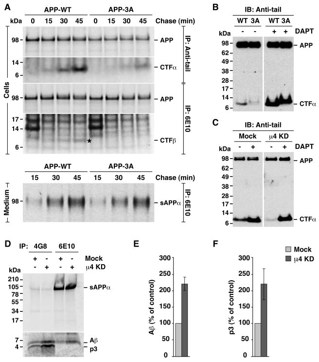
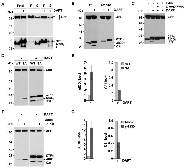
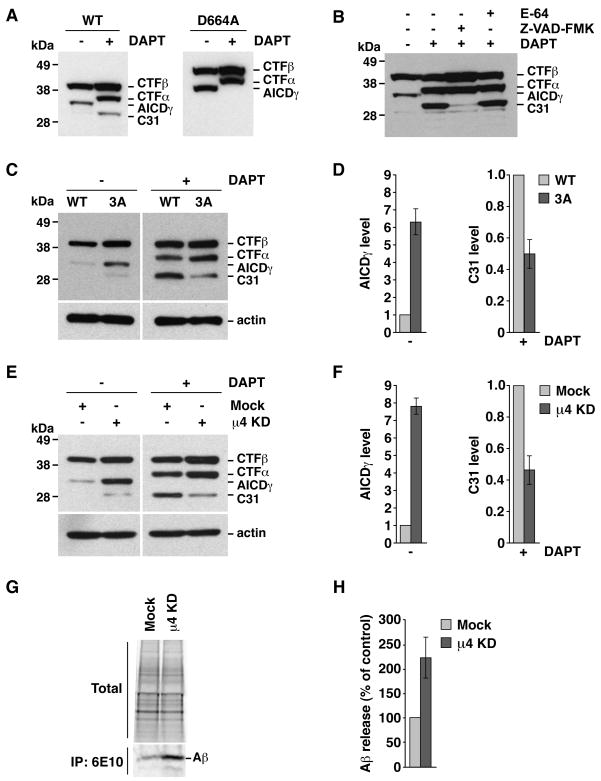
Adaptor protein 4 (AP-4) is the most recently discovered and least well-characterized member of the family of heterotetrameric adaptor protein (AP) complexes that mediate sorting of transmembrane cargo in post-Golgi compartments. Herein, we report the interaction of an YKFFE sequence from the cytosolic tail of the Alzheimer's disease amyloid precursor protein (APP) with the mu4 subunit of AP-4. Biochemical and X-ray crystallographic analyses reveal that the properties of the APP sequence and the location of the binding site on mu4 are distinct from those of other signal-adaptor interactions. Disruption of the APP-AP-4 interaction decreases localization of APP to endosomes and enhances gamma-secretase-catalyzed cleavage of APP to the pathogenic amyloid-beta peptide. These findings demonstrate that APP and AP-4 engage in a distinct type of signal-adaptor interaction that mediates transport of APP from the trans-Golgi network (TGN) to endosomes, thereby reducing amyloidogenic processing of the protein.


Figures

Fig. 1
Fig. 1
(A) Schematic representation of APP indicating its topological domains (TM: transmembrane; N: N -terminus; C: C-terminus), the position of the Aβ peptide, the α, β, γ and caspase cleavage sites, and the fragments produced. Underlined are regions in APP recognized by the antibodies used in this study. (B) Sequence of the APP cytosolic tail indicating residue numbers, two YXXØ motifs (solid underlines), Tyr residues (asterisks), an NPXY-type signal (dashed underline), and key residues for interaction with μ4 (dashed box). (CE and G) Y2H analysis of the interaction of the APP cytosolic tail with μ4. Yeast were co-transformed with plasmids encoding Gal4bd fused to the wild-type or mutant APP constructs indicated on the left, and Gal4ad fused to the adaptor subunits indicated on top of each panel. Mouse p53 fused to Gal4bd and SV40 large T antigen (T Ag) fused to Gal4ad were used as controls. Co-transformed cells were spotted onto His-deficient (−His) or His-containing (+His) plates and incubated at 30°C. (F) ITC of ENPTYKFFEQ peptide (black line and solid circles) or ENPTAKAAEQ peptide (dashed line and open circles) with μ4, and of ENPTYKFFEQ peptide with μ4-R283D (grey line and circles). The Kd and stoichiometry (N) for the μ4-ENPTYKFFEQ interaction are expressed as the mean ± SEM (n=3).
Fig. 2
Fig. 2
Crystal structure of the μ4 C-terminal domain in complex with a peptide signal from APP. (A) Ribbon representation of human μ4 C-terminal domain with subdomain A in blue, subdomain B in red, and the APP peptide (TYKFFEQ; stick model) in yellow. (B) Superposition of μ4 and rat μ2 (with the EGF receptor peptide in magenta; pdb entry 1BW8, Owen and Evans, 1998). In orange (A) is show n a loop that is disordered in μ2. The position of the N- (N, Nμ2 and Nμ4) and C-termini (C) are indicated. (C and D) Model of the AP-4 (C) and structure of the AP-2 core (D; Collins et al. 2002; pdb entry 1GW5) core complexes showing the position of the respective peptide-binding sites and the PIP2–binding site on the AP-2 α subunit. See also Fig. S1.
Fig. 3
Fig. 3
Details and verification of the signal-binding site on μ4. (A) Stick representation of the bound peptide TYKFFEQ (shown with carbon atoms colored yellow) superimposed on a 2Fo-Fc omit electron density map contoured at 0.8 σ. (B) Surface complementarity between the peptide and μ4. Surface colors for residues in contact with the TYKFFEQ peptide are green for hydrophobic, red for acidic, blue for basic, and orange for uncharged polar. (C) Alignments of the sequences containing the signal-binding site in μ4 from different species, and of the homologous μ1A, μ1B, μ2, μ3A and μ3B sequences from H. sapiens. Residues in the signal-binding site in human μ4 that are identical (red), conserved (orange), and non-conserved (gray) are indicated. (D, E) Y2H assays involving co-expression of Gal4ad fused to wild-type or mutant μ4, and Gal4bd fused to the APP tail, were performed as described in the legend to Fig. 1. See also Fig. S2.
Fig. 4
Fig. 4
Redistribution of APP from endosomes to the TGN upon disruption of its interaction with μ4. (A–F) HeLa cells were transfected with plasmids encoding either APP-CFP (WT) (A–C) or APP-CFP carrying the triple mutation, F689A, F690A and E691A (3A) (D–F). (G–L) HeLa cells were mock-transfected (G–I) or transfected twice with siRNA directed to μ4 (J–L), and then re-transfected with a plasmid encoding APP-CFP. Cells were stained for TGN46 and examined by confocal fluorescence microscopy. Merging red and green channels generated the third picture on each row; yellow indicates overlapping localization. Insets show 2X magnifications. Bars: 10 μm.
Fig. 5
Fig. 5
Disruption of the APP-μ4 interaction alters CTF levels and p3/ Aβ secretion. (A) HeLa cells transfected with HA-tagged wild-type APP (APP-WT) or APP with the triple mutation, F689A, F690A and E691A (APP-3A), were labeled for 15 min with [35S]methionine-cysteine and chased for 0–45 min at 37°C. APP species were immunoprecipitated (IP) from cell lysates or culture media with the indicated antibodies. Proteins were analyzed by SDS-PAGE and fluorography. (B–C) Anti-tail immunoblot (IB) analysis of APP and CTF species from cells expressing APP-WT or APP-3A (B), or from mock- or μ4-knockd own (KD) cells expressing APP-WT (C), in the absence (−) or presence (+) of 250 nM γ-secretase inhibitor, DAPT. (D) Mock- or μ4-KD cells expressing APP-WT were labeled for 4 h at 37°C with [35S]methionine-cysteine, and the culture medium was subjected to immunoprecipitation with antibodies 4G8 (recognizing both p3 and Aβ) and 6E10 (recognizing only Aβ). Proteins were analyzed by electrophoresis on Tricine 10–20% acrylamide gradient gels and fluorography. The positions of molecular mass markers are indicated on the left. (E, F) Mean ± SD from three experiments like that in D. Control: mock. See also Fig. S4.
Fig. 6
Fig. 6
Disruption of the APP-μ4 interaction alters AICDγ and C31 levels. All experiments were performed with HeLa cells expressing normal or mutant APP constructs tagged with CFP, incubated in the absence (−) or presence (+) of DAPT, and analyzed by SDS-PAGE and immunoblotting with antibody to GFP, with the variations indicated below. The positions of molecular mass markers and different APP species are indicated. (A) Cells expressing APP-CFP were homogenized to obtain pellet (P) and supernatant (S) fractions. m, mature; i, immature. (B) Cells expressing normal APP-CFP (WT) or APP-CFP carrying a D664A mutation. (C) Cells expressing normal APP-CFP were treated with 100 μM E-64, 200 μM Z-VAD-FMK and / or 250 nM DAPT. (D) Cells expressing normal APP-CFP (WT) or APP-CFP with the F689A, F690A and E691A mutation (3A). (E) Mean ± SD of AICDγ and C31 levels from three experiments such as that shown in D. (F) Mock- or μ4-depleted cells (μ4 KD) expressing APP-CFP. (G) Mean ± SD of AICDγ and C31 levels from three experiments such as that show n in F. See also Fig. S5.
Fig. 7
Fig. 7
Altered processing of CTFβ upon disruption of the YKFFE-μ4 interaction. Experiments were performed as in Fig. 6, with the variations indicated below. The positions of molecular mass markers and different APP species are indicated. (A) Cells expressing normal CTFβ-GFP (WT) or CTFβ-GFP carrying a D664A mutation. (B) Cells expressing normal CTFβ-GFP were treated with 100 μM E-64, 200 μM Z-VAD-FMK and / or 250 nM DAPT. (C) Cells expressing normal CTFβ-GFP (WT) or CTFβ-GFP with the triple mutation, F689A, F690A and E691A (3A). (D) Mean ± SD of AICDγ and C31 levels from three experiments such as that shown in C. (E) Mock- or μ4-depleted cells (μ4 KD) expressing CTFβ-GFP. (F) Mean ± SD of AICDγ and C31 levels from three experiments such as that shown in E. (G) Mock- or μ4-depleted cells expressing normal CTFβ-GFP were labeled for 4 h at 37°C with [35S]methionine-cysteine, and the culture medium was subjected to immunoprecipitation with antibody 6E10 to Aβ. Proteins were analyzed by electrophoresis on Tricine 10–20% acrylamide gradient gels and fluorography. An aliquot of the culture media (total) is shown as loading control. (H) Mean ± SD from four experiments like that in G. Control: mock.

Similar articles

Cited by

References

    1. Aguilar RC, Boehm M, Gorshkova I, Crouch RJ, Tomita K, Saito T, Ohno H, Bonifacino JS. Signal-binding specificity of the mu4 subunit of the adaptor protein complex AP-4. J Biol Chem. 2001;276:13145–13152. - PubMed
    1. Anliker B, Muller U. The functions of mammalian amyloid precursor protein and related amyloid precursor-like proteins. Neurodegener Dis. 2006;3:239–246. - PubMed
    1. Baulac S, LaVoie MJ, Kimberly WT, Strahle J, Wolfe MS, Selkoe DJ, Xia W. Functional gamma-secretase complex assembly in Golgi/trans-Golgi network: interactions among presenilin, nicastrin, Aph1, Pen-2, and gamma-secretase substrates. Neurobiol Dis. 2003;14:194–204. - PubMed
    1. Boehm M, Aguilar RC, Bonifacino JS. Functional and physical interactions of the adaptor protein complex AP-4 with ADP-ribosylation factors (ARFs) Embo J. 2001;20:6265–6276. - PMC - PubMed
    1. Bonifacino JS, Traub LM. Signals for sorting of transmembrane proteins to endosomes and lysosomes. Annu Rev Biochem. 2003;72:395–447. - PubMed

Publication types

MeSH terms

Substances

Associated data