. 2010 Jul;91(1):2-8.
doi: 10.1016/j.exer.2010.03.004. Epub 2010 Mar 15.

Variations in the rheostat model of apoptosis: what studies of retinal ganglion cell death tell us about the functions of the Bcl2 family proteins

Affiliations

Variations in the rheostat model of apoptosis: what studies of retinal ganglion cell death tell us about the functions of the Bcl2 family proteins

Robert W Nickells. Exp Eye Res. 2010 Jul.

Abstract

Studies of the functions of members of the Bcl2 gene family suggested that apoptosis was controlled by a rheostat in which anti-apoptotic proteins like BCL2 bound and sequestered pro-apoptotic proteins like BAX. Our current understanding of these proteins suggests that this is a simplistic model. The new rheostat model predicts that BH3-only peptides act as neutralizing ligands for the anti-apoptotic proteins, thus allowing molecules like BAX to become activated and initiate mitochondrial dysfunction - a critical step in the intrinsic apoptotic program. Studies of retinal ganglion cell apoptosis indicate that a threshold of BAX expression is required for its successful activation, which is independent of the overall concentration of anti-apoptotic proteins in these cells.


Figures

Fig. 1
Fig. 1
Quantitative analysis of BclX and Bax expression in the mouse retina. (A) Quantitative PCR of BclX and Bax transcripts in the retinas of Bax+/+ and Bax+/− mice. BclX mRNA was quantified from Bax+/− animals, but similar levels are detected in animals of all three possible Bax genotypes. In each case, transcript abundance was calculated against a standard curve for each PCR product that was done in concert with the qPCR of the retinal cDNA. Loading of sample template was controlled by the separate quantification of mRNA for the S16 small ribosomal subunit protein. Bax+/− mice express approximately 50% of the amount of Bax mRNA as wild type mice. Alternatively, the same samples exhibit approximately 10 fold higher levels of BclX mRNA. (B) Western blots of Bax+/+ and Bax+/− mice for ACTIN, BCL-X, and BAX protein. Development of the blots was maximized for BCL-X levels, at which point BAX protein is barely, if at all, detectable. In a separate blot, BAX levels were visible using a more sensitive chemiluminescent ECF detection system. Although western blotting technology is limited in its ability to compare quantities of different proteins in the same sample, results like this corroborate quantitative analyses showing much greater levels of BclX mRNA in the retina.
Fig. 2
Fig. 2
Summary diagram of the proposed mechanism of BAX activation and function. (A) In most cells, BAX exists as a latent globular protein already present in the cytosol. The folded structure hides a hydrophobic cleft that is partially composed of the BH3 homology domain (green box), and a putative C-terminal domain, which has structural similarity to a mitochondrial targeting sequence (red box). The process of BAX activation begins with a change in protein conformation, which exposes the hydrophobic cleft, the N-terminus (containing the antigenic site for the 6A7 monoclonal antibody), and the mitochondrial targeting sequence. Recent studies indicate that BAX unfolding is initiated by separate peptides similar to the BIM BH3 domain, although this interaction is not directly with the BAX BH3 domain. (B) Either in concert with the unfolding step, or as a consequence of it, BAX becomes integrated into the mitochondrial outer membrane (MOM). Studies on other proteins containing mitochondrial targeting sequences, suggest that this domain interacts with some chaperone that helps direct these proteins to the appropriate organelles. Currently, there is no direct evidence that BAX interacts with a cellular chaperone, but BAX constructs missing this domain are not able to translocate to mitochondria in the absence of wild type BAX proteins. (C) Once some BAX proteins become inserted into the MOM, they appear to act as a sink for other BAX molecules. In this secondary recruitment step, truncated BAX proteins lacking the translocation domain are able to insert into the MOM, suggesting that they are activated by direct interaction with exposed BH3 domains of BAX molecules already in the membrane. As BAX molecules aggregate in the MOM, they potentially create pore structures that facilitate the release of cytochrome c and downstream events of the apoptotic cascade.
Fig. 3
Fig. 3
Summary of the relationship between retinal ganglion cell apoptosis and Bax expression in two lines of mice. Original data for this model is given in Semaan et al (Semaan et al., 2010). Experiments were conducted on two strains (129B6 and DBA/2J), which carried a mutant allele of Bax. Left eyes of mice were subjected to optic nerve crush (Li et al., 1999), and cells in the retinal ganglion cell layer were counted in both eyes, two weeks after the crush surgery. Bax−/− ganglion cells in both lines exhibited complete resistance to optic nerve crush (white zone), while wild type mice from both lines exhibited comparable levels of cell loss (grey zone). DBA/2JBax+/− mice, exhibited complete resistance, similar to knock-out mice. Conversely, 126B6Bax+/− mice exhibited normal cell loss. Quantitative evaluation of both transcripts and protein in these revealed that DBA/2J animals carried approximately half of the BAX present in 129B6 mice. Comparatively, transgenic mice overexpressing Bax under the control of the Neuron specific enolase promoter (NseBax) show normal kinetics of neuronal apoptosis (Bernard et al., 1998). These data suggest that the commitment to cell death is an all-or-none phenomenon, and that BAX levels (stippled box) must be at a certain threshold to reach this committed step.
Fig. 4
Fig. 4
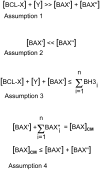
Mathematical assumptions governing variations in the rheostat model. In this series of equations, important aspects of the different levels of members of the Bcl2 gene family proteins are considered for their roles in blocking apoptosis and/or in the activation of the intrinsic cell death program. Variables are given as either cellular concentrations (written in square brackets) or as absolute numbers of molecules, in which each variable is written in summation form. The variables are: BCL-X = the principal anti-apoptotic protein expressed in retinal ganglion cells, Y = other molecules of the Bcl2 gene family with anti-apoptotic properties. This may include, but not be limited to, molecules such as BCL2, MCL1, BCL-W, etc. BAX′ = molecules of BAX that are initially converted from a globular soluble structure to become inserted in the mitochondrial outer membrane. For the purposes of this argument, this is considered the primary BAX activating event, and probably requires interaction with one or more members of the BH3-only peptide family. BAX″ = all other latent BAX molecules that are present in the cytosol, but are not activated by BH3-only peptide interactions. Instead, these molecules become the pool of BAX proteins that are secondarily activated after the unfolding and insertion of BAX′ molecules. BAXCM = the critical mass of BAX molecules that must be assembled as oligomers in the mitochondrial membrane. This is a hypothetical variable and makes the assumption that functional destabilization of the mitochondrial membrane requires a specific density of BAX proteins to assemble. BH3 = BH3-only peptides that become activated during the course of apoptosis of ganglion cell somas. Assumption 1. This equation summarizes the probable association of BAX molecules and anti-apoptotic molecules present in living, healthy, ganglion cells. Semi-quantitative western blots to measure protein levels, and quantification of transcripts using real-time PCR suggest that just BCL-X alone is several fold more abundant than BAX levels in ganglion cells. Although BAX must become activated in order to execute the cell death program, the excess of antagonistic BCL-X likely prevents “accidental” activation events, which may be one reason why BCL-X is already localized to the surface of organellar membranes. Assumption 2. This equation summarizes the hypothetical assumption that BAX activation occurs in two stages. The first stage requires only minimal initial activation of BAX (BAX′) by interaction of globular BAX with a BH3-only protein. Once unfolded, BAX′ is able to both insert into the mitochondrial outer membrane, and act as a sink to activate other molecules of globular BAX in the cytosol (BAX″). This event, by itself, is probably not sufficient to elicit cell death, but is likely sufficient to initiate recruitment of other BAX proteins. Assumption 3. This equation summarizes the critical event leading to the activation of BAX′ molecules, and is essentially the core element of the modern rheostat model. In this model, enough BH3-only proteins must be recruited to inactivate all anti-apoptotic proteins present in a living cell ([BCL-X] + [Y]) as well as still be available to activate BAX′. Equilibrium effects, in which the binding of BH3-only proteins is favored for some Bcl2 family proteins over others, probably requires and excess of BH3-only peptides (n) in order to form partners with all the relevant variables in this equation. Assumption 4. These two equations summarize the threshold requirements for BAX in order to form functional oligomers in the mitochondrial outer membrane. First, a critical mass of activated BAX is required for oligomer formation. The critical mass is composed of BAX′ molecules, and some portion (n) of BAX″ molecules. It is likely, however, that cells generally contain enough latent BAX molecules to achieve BAXCM, and cells with large excesses of BAX do not exhibit a greater cell death response once critical mass is established. Cells with BAX content below the critical mass threshold are resistant to apoptosis, however. In this model, I assume that [BAX′] ≪ [BAXCM], therefore, subthreshold levels of BAX probably affect the efficiency of secondary recruitment of BAX″ molecules to the mitochondria, thus delaying the formation of BAXCM. Evidence for this is the very slow activation of apoptosis over several months in Bax+/− retinal ganglion cells that are initially resistant to optic nerve crush (Semaan et al., 2010).

Similar articles

Cited by

References

    1. Adams JM, Cory S. The bcl-2 protein family: arbiters of cell survival. Science. 1998;281:1322–1326. - PubMed
    1. Adams JM, Cory S. The Bcl-2 apoptotic switch in cancer development and therapy. Oncogene. 2007;26:1324–1337. - PMC - PubMed
    1. Antonsson B, Conti F, Ciavatta A, Montessuit S, Lewis S, Martinou I, Bernasconi L, Bernard A, Mermod JJ, Mazzei G, Maundrell K, Gambale F, Sadoul R, Martinou JC. Inhibition of Bax channel-forming activity by Bcl-2. Science. 1997;277:370–372. - PubMed
    1. Antonsson B, Montessuit S, Lauper S, Eskes R, Martinou JC. Bax oligomerization is required for channel-forming activity in liposomes and to trigger cytochrome c release from mitochondria. Biochem. 2000;345:271–278. - PMC - PubMed
    1. Antonsson B, Montessuit S, Sanchez B, Martinou JC. Bax is present as a high molecular weight oligomer/complex in the mitochondrial membrane of apoptotic cells. J Biol Chem. 2001;276:11615–11623. - PubMed

Publication types

MeSH terms