. 2010 Jun;138(7):2509-18, 2518.e1.
doi: 10.1053/j.gastro.2010.03.008. Epub 2010 Mar 12.

Hepatitis C virus regulates transforming growth factor beta1 production through the generation of reactive oxygen species in a nuclear factor kappaB-dependent manner

Affiliations

Hepatitis C virus regulates transforming growth factor beta1 production through the generation of reactive oxygen species in a nuclear factor kappaB-dependent manner

Wenyu Lin et al. Gastroenterology. 2010 Jun.

Abstract

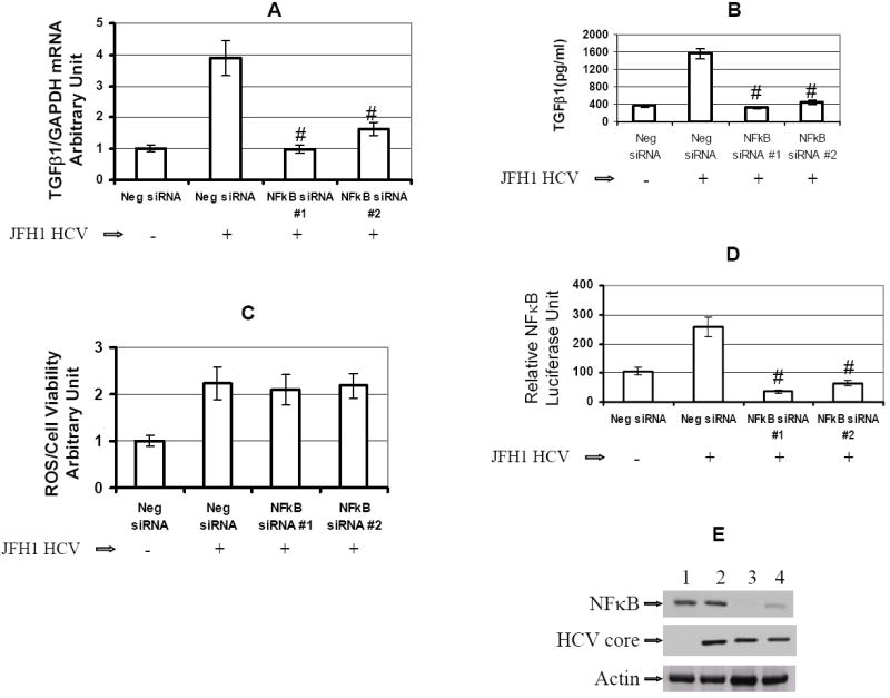
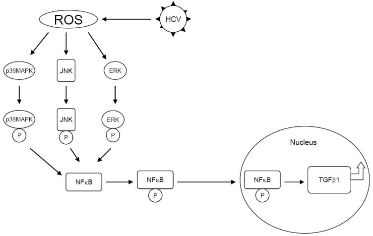
Background & aims: The generation of oxidative stress and transforming growth factor beta1 (TGF-beta1) production play important roles in liver fibrogenesis. We have previously shown that hepatitis C virus (HCV) increases hepatocyte TGF-beta1 expression. However, the mechanisms by which this induction occurs have not been well studied. We explored the possibility that HCV infection regulates TGF-beta1 expression through the generation of reactive oxygen species (ROS), which act through > or =1 of the p38 mitogen-activated protein kinase (MAPK), extracellular signal-regulated kinase (ERK), c-Jun N-terminal kinase (JNK), and nuclear factor kappaB (NFkappaB) signaling pathways to induce TGF-beta1 expression.

Methods: We used small molecule inhibitors and short interfering RNAs to knock down these pathways to study the mechanism by which HCV regulates TGF-beta1 production in the infectious JFH1 model.

Results: We demonstrated that HCV induces ROS and TGF-beta1 expression. We further found that JFH1 induces the phosphorylation of p38MAPK, JNK, ERK, and NFkappaB. We also found that HCV-mediated TGF-beta1 enhancement occurs through a ROS-induced and p38 MAPK, JNK, ERK1/2, NFkappaB-dependent pathway.

Conclusions: These findings provide further evidence to support the hypothesis that HCV enhances hepatic fibrosis progression through the generation of ROS and induction of TGF-beta1. Strategies to limit the viral induction of oxidative stress appear to be warranted to inhibit fibrogenesis.


Conflict of interest statement

There is no conflict to disclose for the authors.

Figures

Figure 1
Figure 1. HCV increases TGF-β1 mRNA and ROS production in JFH1-infected Huh7.5.1 cells
Figure 1A. JFH1 HCV induces ROS production. ROS level was normalized by cell viability to calculate the ROS/Cell Viability Arbitrary Unit. Empty bar: Huh7.5.1 cell, Gray bar: JFH1 cell. (n=4, P=0.001) Figure 1B. JFH1 increased ROS fluorescence. ROS fluorescent images in Huh7.5.1 and JFH1 live cells. Figure 1C. JFH1 HCV increases TGF-β1 expression. TGF-β1 level was normalized to GAPDH level to calculate the TGF-β1/GAPDH arbitrary unit. Empty bar: huh7.5.1 cell, Gray bar: JFH1 cell. (n=4, P=0.007). Figure 1D. Western blot for HCV NS5A and HCV core. HCV NS5A, HCV core, and actin proteins levels were detected by Western blots. Lane #1: Huh7.5.1 cells; Lane#2: JFH1 cells.
Figure 2
Figure 2. The ROS inhibitor DPI blocks HCV upregulation of TGF-β1
The inhibitors used including DPI, SB, SP, U0126, or LY. 1% DMSO was used as a negative control. Figure 2A. DPI blocked JFH1 HCV mediated ROS generation. ROS level was normalized by cell viability to calculate the ROS/Cell Viability Arbitrary Unit. DPI completely blocked the HCV induced ROS production when compared to JFH1 in DMSO (P<0.001, n=4). Figure 2B. DPI inhibited JFH1 HCV mediated TGF-β1 expression. TGF-β1 level was normalized to GAPDH level to calculate the TGF-β1/GAPDH arbitrary unit. DPI completely blocked the HCV stimulated TGF-β1 expression (P=0.002). SB, SP, or U0126 partially reduced TGF-β1 production by 29.5% (P=0.08), 32.2% (P=0.07), and 27.5% (P=0.1), respectively compare to JFH1 in DMSO (n=4). In contrast, LY had no effect on TGF-β1 expression. Figure 2C. The ROS inhibitor DPI blocked phosphorylation of p38 MAPK, JNK, ERK. JFH1 HCV activated the phosphorylation of p38 MAPK, p42 ERK, and JNK (lane #2). ROS inhibitor DPI blocks phosphorylation of p38 MAPK, JNK, ERK to levels comparable to those seen with their specific inhibitors (Lane #3). Antibody to PI3K was directed against unphosphorylated protein. We found that HCV does not activate PI3K phosphorylation (data not shown). Lane#1: Huh7.5.1+DMSO; Lane#2 JFH1+DMSO; Lane#3 JFH1+DPI; Lane#4 JFH1+SB; #5 JFH1+SP; #6 JFH1+U0126; #7 JFH1+LY.
Figure 3
Figure 3. Effects of siRNA to p38 MAPK, JNK, ERK, or PI3K on ROS production and TGF-β1 expression
Figure 3A. siRNA to p38 MAPK, JNK, ERK, or PI3K do not effect JFH1 HCV stimulated ROS production. JFH1 cell increased ROS production by over two-fold compared to Huh7.5.1 cell. However, siRNA to p38 MAPK, JNK, ERK, PI3K had not effect on ROS production in JFH1 infected cells. Figure 3B. siRNA to p38 MAPK, JNK, ERK, partially reduced JFH1 HCV activated TGF-β1 mRNA expression. HCV infection enhanced TGF-β1 expression by over 3 fold in JFH1 cells compared to Huh7.5.1 cells. The siRNA to p38 MAPK, JNK, or ERK partially inhibited TGFβ1 mRNA expression enhancement in JFH1 cells by 45.9% (P=0.035), 40.8% (P=0.070), and 41.4% (P=0.056), respectively, when compared to TGF-β1 mRNA in Neg siRNA in JFH1 (n=4). In contrast, PI3K or Neg siRNA had not effect on TGFβ1 expression in JFH1 cells. Figure 3C. siRNA to p38 MAPK, JNK, ERK significantly inhibited JFH1 HCV-activated TGF-β1 cytokines. TGF-β1 levels in supernatants were measured according to Methods. siRNA to p38 MAPK, JNK, or ERK each significantly reduced TGF-β1 cytokine levels to 1105±88 pg/ml (P=0.015), 1102±95 pg/ml (P=0.016), and 1073±105 pg/ml (P=0.016), respectively, when compared to TGF-β1 levels in the presence of Neg siRNA in JFH1-infected cells (1571±111 pg/ml, n=4). Figure 3D. siRNA to p38 MAPK, ERK, or JNK reduced HCV mediated NFκB phosphorylation. siRNA to p38 MAPK, JNK, ERK, or PI3K knocked down the correspondent protein expression. siRNA to p38 MAPK, JNK, ERK reduced HCV mediated NFκB activation. In contrast, PI3K siRNA has not effect on NFκB phosphorylation. Lane#1 Huh7.5.1, #2 JFH1 + Neg siRNA, #3JFH1+ p38 MAPK siRNA, #4 JFH1+ JNK siRNA, #5 JFH1+ ERK siRNA, #6 JFH1+ PI3K siRNA.
Figure 4
Figure 4. The combination of siRNA to p38MAPK and ERK further reduced TGF-β1 production
Figure 4A. The combination of siRNA to p38MAPK and siRNA to ERK cooperatively reduced TGF-β1 mRNA expression. TGF-β1 mRNA expression in JFH1-infected cells were partially inhibited by siRNAs to p38 MAPK (P=0.031) or ERK (P=0.046) compared to Neg siRNA. The combination of siRNA to p38MAPK and siRNA to ERK additively reduced TGF-β1 mRNA expression in JFH1 cell (P<0.001). Figure 4B. The combination of siRNAs to p38MAPK and ERK significantly inhibited JFH1 HCV-activated TGF-β1 cytokines. TGF-β1 levels in supernatants were measured using the human TGF-β1 ELISA Kit. The combination of siRNAs to p38MAPK and ERK significantly inhibited TGF-β1 cytokine production (596±73 pg/ml, P<0.001) when compared for Neg siRNA in JFH1 cells (1571±111 pg/ml, n=4). Figure 4C. The combination of siRNA to p38MAPK and siRNA to ERK further inhibited HCV-mediated NF-κB phosphorylation. Western blot confirmed that siRNA to p38 MAPK or ERK knocked down the corresponding protein expression. The combination of siRNA to p38 MAPK and ERK further reduced HCV-mediated NFκB activation. Lane#1 Huh7.5.1+ Neg siRNA, #2 JFH1 + Neg siRNA, #3 JFH1+ p38 MAPK siRNA, #4 JFH1+ ERK siRNA, #5 JFH1+ p38 MAPK siRNA+ ERK siRNA.
Figure 5
Figure 5. Effects of ROS inhibitor on NF-κB signaling pathway
An NFκB promoter construct expressing firefly (pNFκB-Luc) and the control construct pRL-TK expressing Renilla luciferase were transfected into cells for 24 hours. The transfected Huh7.5.1 cells or JFH1 cells were incubated with 20 μM different inhibitors for 14 hours. The inhibitors used included DPI, SB, SP, AQ, U0126, or LY. 1% DMSO was used as a negative control. Designations are the same in Figure 5A and B. Figure 5A. DPI blocks HCV-activated NFκB signaling. Relative luciferase activity (RLA) was normalized by dividing the firefly luciferase value by the Renilla luciferase value. HCV increased NFκB promoter signaling by over two fold. DPI and AQ completely blocked NFκB signaling. SB, SP, and U0126 reduced NFκB signaling by 82.9%, 76.7%, and 52.1%, respectively. In contrast, LY 294002 had no effect on NFκB signaling. Figure 5B. DPI blocked HCV-mediated NFκB phosphorylation. HCV activated NFκB phosphorylation in JFH1 cells compared to Huh7.5.1 cells. DPI and AQ blocked NFκB phosphorylation. SB, SP, or U0126 partially reduced NFκB phosphorylation. In contrast, LY had no effect on NFκB phosphorylation. Lane#1 Huh7.5.1, #2 JFH1, Lane#3 JFH1+DPI; Lane#4 JFH1+SB; #5 JFH1+SP; #6 JFH1+AQ; #7 JFH1+U0126; #8 JFH1+LY.
Figure 6
Figure 6. Effects of NFκB siRNA on ROS production and TGF-β1 expression
The siRNAs were transfected into Huh7.5.1 cells or JFH1 cells in 96-well plate for 72 hr. The siRNAs used for gene knocked down included Neg siRNA, NFκB siRNA#1, or NFκB siRNA#2. ROS generation and TGF-β1 expression were assessed in these siRNA transfected cells. Figure 6A. NFκB siRNA reduced JFH1 HCV activated TGF-β1 mRNA expression. TGF-β1 expression was assessed in these siRNA transfected cells. HCV enhanced TGF-β1 expression by over 3 fold in JFH1-infected cells compared to Huh7.5.1 cells. NFκB specific siRNA#1 or siRNA#2 inhibited enhancement of TGF-β1 expression in JFH1 cells. Figure 6B. NFκB siRNA significantly inhibited HCV-activated TGF-β1 cytokine production. TGF-β1 cytokine levels in supernatants were measured by ELISA. Both NFκB-specific siRNA #1 and siRNA #2 significantly reduced JFH1-induced TGF-β1 cytokine levels to 325±30 pg/ml (P<0.001), and 440±48 pg/ml (P<0.001), respectively, when compared to Neg siRNA in JFH1-infected cells (1571±111 pg/ml, n=4). Figure 6C. NFκB siRNA had no effect on JFH1 HCV stimulated ROS production. ROS generation was measured in these siRNA transfected cells. HCV increased ROS production by over 2 fold in JFH1 cells. However, NFκB siRNA#1 or siRNA#2 had not effect on ROS production in JFH1 cells. Figure 6D. NFκB siRNA inhibited NFκB signaling. To monitor the effect of NFκB siRNA on NFκB signaling, at 48 hours after siRNA transfection, NFκB promoter construct expressing firefly (pNFκB-Luc) and construct pRL-TK expressing Renilla luciferase were transfected into these siRNA transfected cells. Cell lysates were harvested for the luciferase assay after 24 hours of NFκB promoter transfection. Firefly and Renilla dual-luciferase activity was measured. RLA was normalized by dividing the firefly luciferase value by the Renilla luciferase value. Both NFκB siRNA #1 and siRNA #2 inhibited the HCV mediated NFκB signaling. Figure 6E. NFκB siRNA knocked down NFκB protein expression. NFκB siRNA#1 or siRNA#2 knocked down the NFκB protein expression. Lane#1 Huh7.5.1+ Neg siRNA, #2 JFH1 + Neg siRNA, #3 NFκB siRNA #1, #4 NFκB siRNA #2.
Figure 7
Figure 7. Schematic model of possible pathway of HCV increases TGF-β1 expression through generation of ROS and NFκB activation
HCV infection induces ROS generation. ROS activate the phosphorylation of p38 MAPK, JNK, and ERK. The phosphorylated p38 MAPK, JNK, and p42/44 MEK subsequently activate the phosphorylation of NFκB. The activated NFκB translocates to the nucleus to upregulate large amount of cytokine genes including TGF-β1 production.

References

    1. Alter MJ. Epidemiology of viral hepatitis and HIV co-infection. J Hepatol. 2006;44:S6–9. - PubMed
    1. Lauer GM, Walker BD. Hepatitis C virus infection. N Engl J Med. 2001;345:41–52. - PubMed
    1. Blackard JT, Kang M, Sherman KE, Koziel MJ, Peters MG, Chung RT. Effects of HCV treatment on cytokine expression during HCV/HIV coinfection. J Interferon Cytokine Res. 2006;26:834–8. - PMC - PubMed
    1. Bataller R, Paik YH, Lindquist JN, Lemasters JJ, Brenner DA. Hepatitis C virus core and nonstructural proteins induce fibrogenic effects in hepatic stellate cells. Gastroenterology. 2004;126:529–40. - PubMed
    1. Lin W, Weinberg EM, Tai AW, Peng LF, Brockman MA, Kim KA, Kim SS, Borges CB, Shao RX, Chung RT. HIV increases HCV replication in a TGF-beta1-dependent manner. Gastroenterology. 2008;134:803–11. - PubMed

Publication types

MeSH terms

Substances