Review
. 2010 Aug;1800(8):824-33.
doi: 10.1016/j.bbagen.2010.03.004. Epub 2010 Mar 15.

Insect ferritins: Typical or atypical?

Affiliations
Review

Insect ferritins: Typical or atypical?

Daphne Q D Pham et al. Biochim Biophys Acta. 2010 Aug.

Abstract

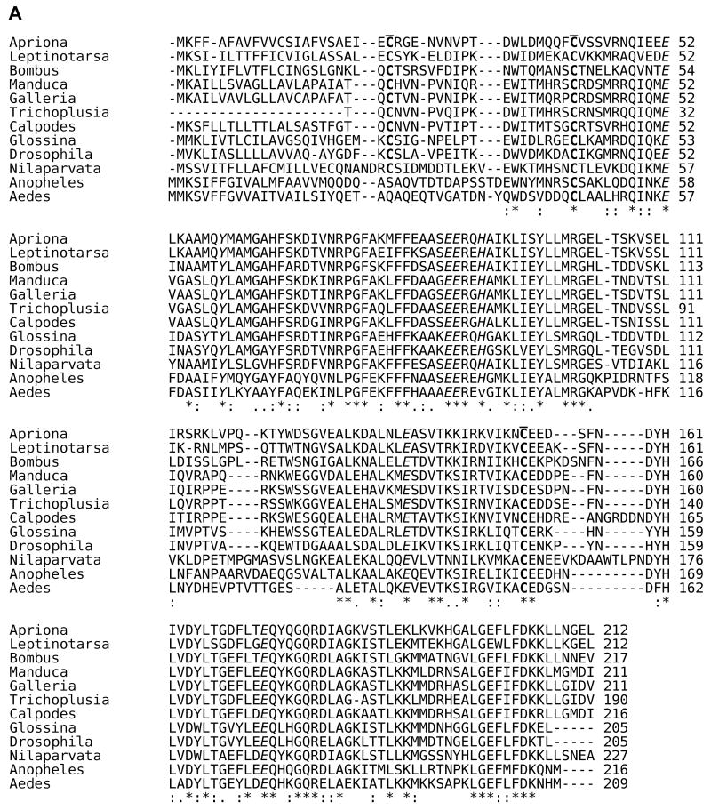
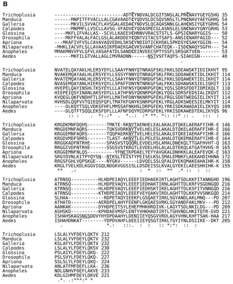
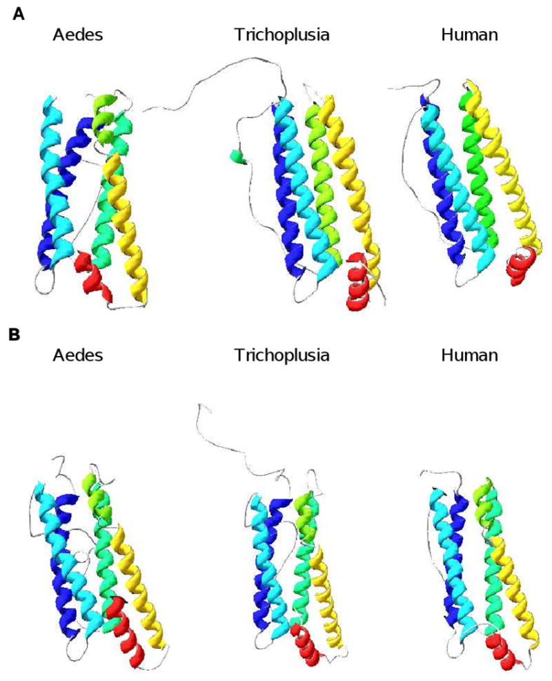
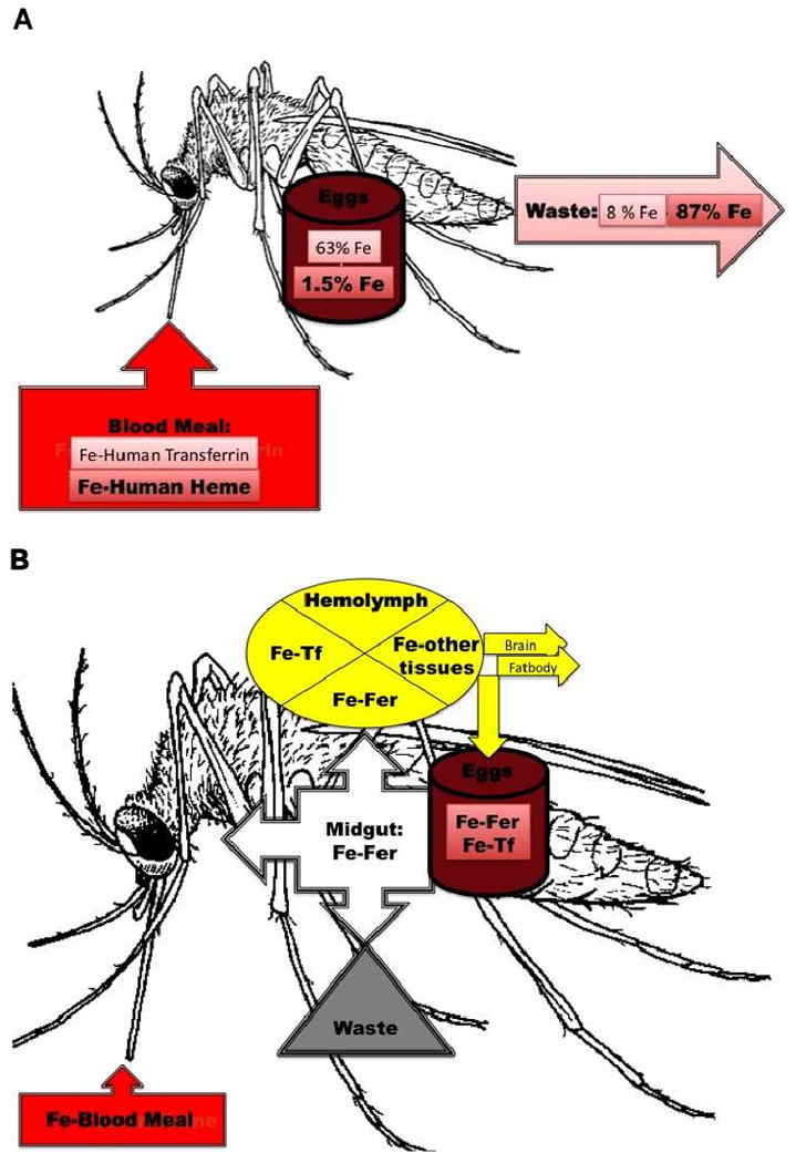
Insects transmit millions of cases of disease each year, and cost millions of dollars in agricultural losses. The control of insect-borne diseases is vital for numerous developing countries, and the management of agricultural insect pests is a very serious business for developed countries. Control methods should target insect-specific traits in order to avoid non-target effects, especially in mammals. Since insect cells have had a billion years of evolutionary divergence from those of vertebrates, they differ in many ways that might be promising for the insect control field-especially, in iron metabolism because current studies have indicated that significant differences exist between insect and mammalian systems. Insect iron metabolism differs from that of vertebrates in the following respects. Insect ferritins have a heavier mass than mammalian ferritins. Unlike their mammalian counterparts, the insect ferritin subunits are often glycosylated and are synthesized with a signal peptide. The crystal structure of insect ferritin also shows a tetrahedral symmetry consisting of 12 heavy chain and 12 light chain subunits in contrast to that of mammalian ferritin that exhibits an octahedral symmetry made of 24 heavy chain and 24 light chain subunits. Insect ferritins associate primarily with the vacuolar system and serve as iron transporters-quite the opposite of the mammalian ferritins, which are mainly cytoplasmic and serve as iron storage proteins. This review will discuss these differences.


Figures

Fig. 1
Fig. 1. Amino acid sequence alignment for insect HCH and LCH subunits
The signal sequences for both the HCH and LCH T. ni subunits are removed to maintain the same numerical assignments as cited in the original structural work [31]. * = identical residues; : = conserved residues; . = semi-conserved residues. The sequence alignments were performed using Clustal 2.0.11 multiple sequence alignment at http://www.ebi.ac.uk/Tools/clustalw2/index.html with known insect HCH and LCH sequences deposited at the National Center for Biotechnology Information (http://www.ncbi.nlm.nih.gov/). A. Sequence alignment for insect HCH subunits. C residues involved in inter- and intrasubunit disulside bonds are bolded and overlined; amino acid residues in ferroxidase center are italicized; amino acid residues engaged in the salt bridges and pi-cation interactions are shaded in grey. Sequences are from Manduca sexta (hawkmoth, Lepidoptera) [19], Calpodes ethlius (skipper butterfly, Lepidoptera) [21], Galleria mellonella (wax moth, Lepidoptera) [23], Nilaparvata lugens (plant hopper, Hemiptera) [22], Aedes aegypti (yellow fever mosquito, Diptera) [18], Drosophila melanogaster (fruit fly, Diptera) [20], Anopheles gambiae (malaria mosquito, Diptera) [80], Apriona germari (long horned beetle, Coleoptera) [24], Leptinotarsa decemlineata (Colorado potato beetle, Coleoptera [25], Glossina morsitans morsitans (tsetse fly, Diptera) [15], Bombus ignitus (bumble bee, Hymenoptera), [16, 17] and Trichoplusia ni (cabbage looper, Lepidoptera) [31]. B. Sequence alignment for insect LCH subunits. C residues and amino acid residues engaged in the salt bridges and pi-cation interactions are represented as described in (A). Putative N-glycosylation sites (N-X-S/T) are underlined. LCH subunit sequences are from the same species and references cited above; no LCH sequence was reported for B. ignitus.
Fig. 2
Fig. 2. Molecular modeling for insect HCH and LCH subunits
The molecular modeling [81-83] was performed at Swiss-Model website (http://swissmodel.expasy.org). A. Structures of heavy chains. Aedes = A. aegypti; Trichoplusia = T. ni; human = Homo sapiens. Helix A = blue; helix B = magenta; helix C = green; helix D = yellow; helix E = red. Color coding: blue = N-terminus; red = C-terminus. Arrows = C-terminus of domain. B. Structures of light chains. Abbreviations and colors are as described in A.
Fig. 3
Fig. 3. Iron metabolism in mosquitoes. A. Main route for iron absorption
Fe-Human Transferrin = iron from human transferrin in the blood meal; Fe-Human Heme = iron from human hemoglobin in the blood meal. Fe (not bolded) = % of Fe-Human Transferrin deposited in eggs or going to waste; Fe (bolded) = % of Fe-Human Heme deposited in eggs or going to waste. B. Proteins involved in iron transfer to different tissues. Fe = iron; Fer = insect ferritin; Tf = insect transferrin.

References

    1. Andrews NC. Forging a field: the golden age of iron biology. Blood. 2008;112:219–230. - PMC - PubMed
    1. Arosio P, Ingrassia R, Cavadini P. Ferritins: A family of molecules for iron storage, antioxidation and more. Biochim Biophys Acta. 2009;1790:589–599. - PubMed
    1. Leimberg JM, Prus E, Link G, Fibach E, Konijn AM. Iron-chelator complexes as iron sources for early developing human erythroid precursors. Transl Res. 2008;151:88–96. - PubMed
    1. Fisher J, Devraj K, Ingram J, Slagle-Webb B, Madhankumar AB, Liu X, Klinger M, Simpson IA, Connor JR. Ferritin: a novel mechanism for delivery of iron to the brain and other organs. Am J Physiol Cell Physiol. 2007;293:C641–649. - PubMed
    1. Levi S, Arosio P. Mitochondrial ferritin. Int J Biochem Cell Biol. 2004;36:1887–1889. - PubMed

Publication types

LinkOut - more resources